
Diagnostic value of 18F-fluorodeoxyglucose positron emission computed tomography, magnetic resonance imaging, and plasma EBV levels on local recurrence of nasopharyngeal carcinoma
-
摘要:
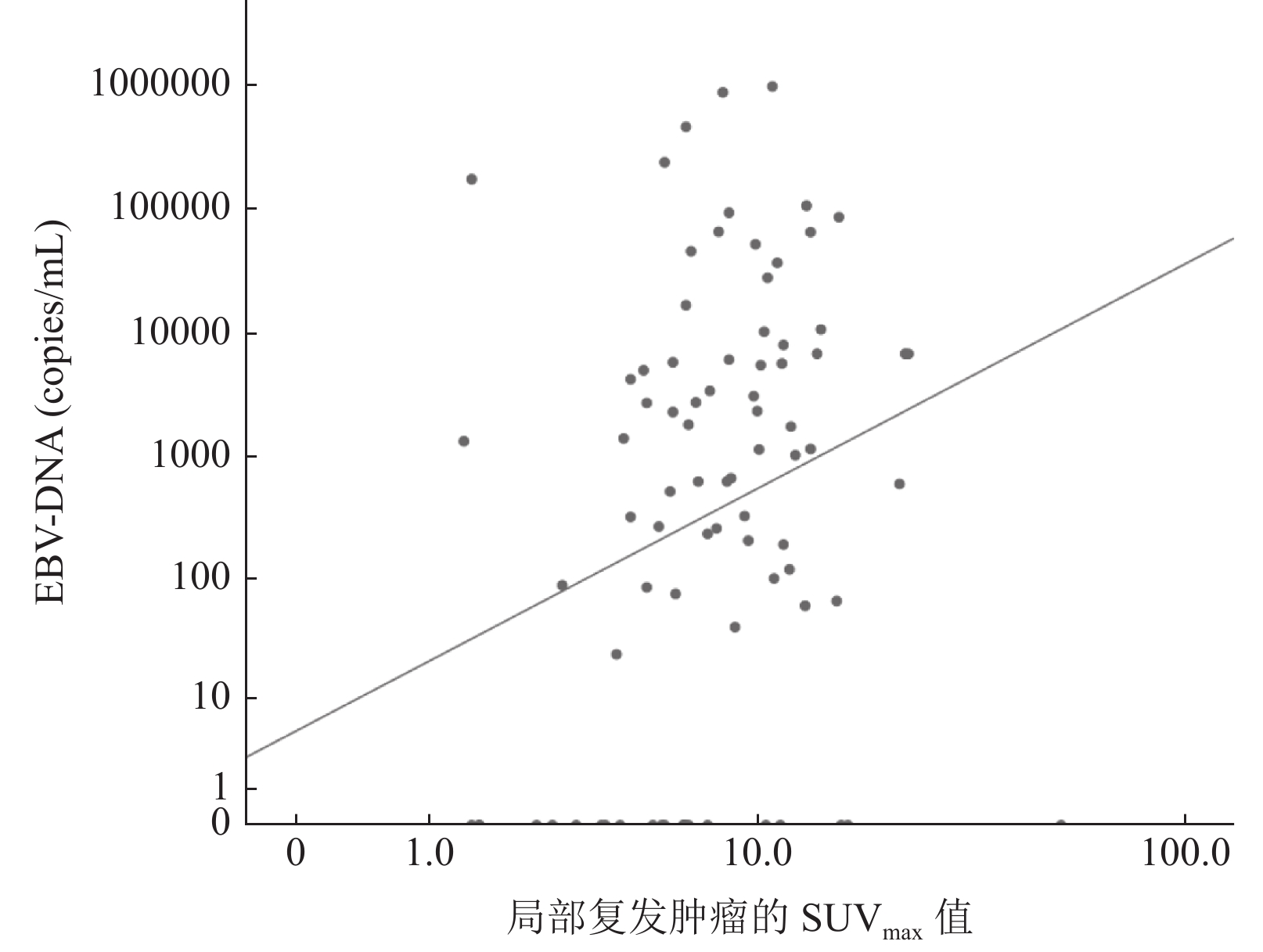
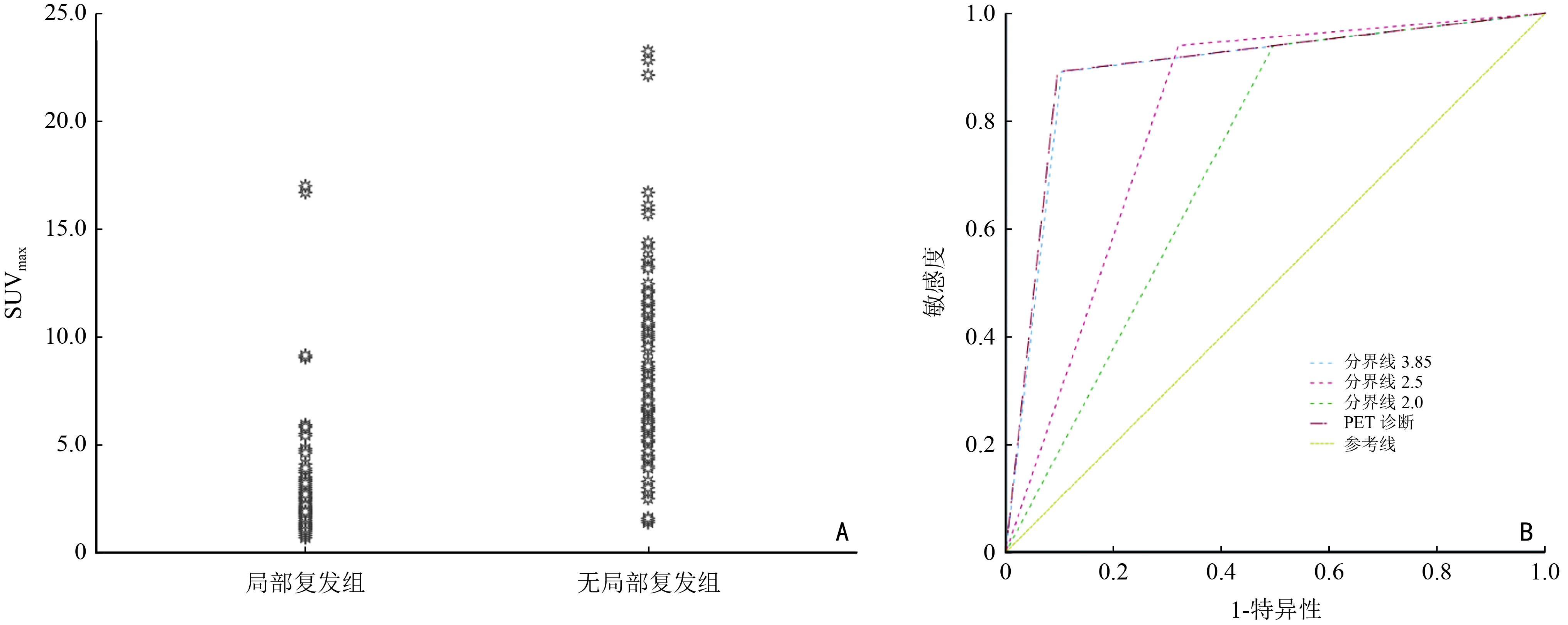
目的 比较18F-FDG PET/CT、MRI在鼻咽癌局部复发和放疗后改变的鉴别诊断中的应用价值,并确定更合适的SUVmax诊断阈值,探索血浆EBV-DNA在发现鼻咽癌复发转移方面的意义。 方法 选取2015年1月至2020年2月期间放疗结束至少6个月后在福建省肿瘤医院进行复查的鼻咽癌患者208例,均进行MRI、PET/CT及EB病毒DNA检查(均在1个月内完成)。以活检病理结果或影像学的密切随访(至少1年)作为诊断鼻咽癌是否局部复发的标准,对患者的影像检查结果及相关参数进行比较分析。 结果 共有83例患者确诊为鼻咽癌局部复发,125例确诊为无局部复发,后者中77例伴有区域复发和(或)远处转移。PET/CT和MRI对鼻咽癌局部复发的诊断敏感性、特异性、准确性分别为89.2% vs. 67.5%,90.4% vs. 92.0%,89.9% vs. 82.2%。当诊断临界值SUVmax=3.85时,PET/CT的诊断准确性最高,与SUVmax=2.5相比,特异性(89.6% vs. 68.0%)和准确性(89.4% vs. 78.4%)均有显著提升。对纳入患者中治疗失败患者的EB病毒DNA分析发现,局部复发患者的血浆EBV-DNA检测敏感性低于区域复发或远处转移患者。局部复发肿瘤患者的SUVmax值与血浆EBV-DNA存在相关性。 结论 18F-FDG PET/CT对鼻咽癌局部复发和放疗后改变的鉴别诊断效能优于MRI。SUVmax诊断阈值设为3.85时可获得更好的诊断效能。血浆EBV-DNA检测在鼻咽癌区域复发和远处转移方面较灵敏,局部复发者的阳性率并不高,对这部分患者的诊断价值仅提供参考。 -
关键词:
- 鼻咽癌 /
- 局部肿瘤复发 /
- 核磁共振成像 /
- 正电子发射计算机断层显像 /
- EB病毒感染
Abstract:Objective To compare the application value of 18F-fluorodeoxyglucose positron emission computed tomography (18F-FDG PET/CT) and magnetic resonance imaging (MRI) in the differential diagnosis of local recurrence and post-radiotherapy changes in nasopharyngeal carcinoma (NPC); to determine a more appropriate diagnostic threshold for maximum standardized uptake value (SUVmax); and to explore the significance of plasma EBV-DNA in detecting recurrent metastases in NPC. Methods Patients with NPC, who underwent review at Fujian Cancer Hospital between January 2015, and February 2020, 6 months after the end of radiotherapy, underwent MRI, PET/CT, and EBV DNA examinations (all within a month). Close follow-up (at least 1 year) of their biopsy pathology results or imaging was used as basis to verify whether NPC was locally recurrent. The patients' imaging findings and related parameters were compared and analyzed. Results A total of 83 patients were diagnosed with a local recurrence of NPC. Besides, 125 were diagnosed without local recurrence. Seventy-seven of the patients without local recurrence had a regional recurrence and/or distant metastases. The sensitivity, specificity, and accuracy of PET/CT and MRI for local recurrence of NPC were 89.2% vs. 67.5%, 90.4% vs. 92.0%, and 89.9% vs. 82.2%, respectively. The diagnostic accuracy of PET/CT was highest when the diagnostic threshold SUVmax=3.85. The specificity (89.6% vs. 68.0%) and accuracy (89.4% vs. 78.4%) significantly improved compared with a SUVmax=2.5. Analysis of EBV-DNA from patients with treatment failure revealed that plasma EBV-DNA detection sensitivity was lower in patients with local recurrence than in patients with regional recurrence or distant metastases. This highlights the correlation between SUVmax values and plasma EBV-DNA in patients with locally recurrent tumors. Conclusions 18F-FDG PET/CT is more effective than MRI for the differential diagnosis of local recurrence and post-radiation changes in NPC. Better diagnostic efficacy is obtained when the SUVmax diagnostic threshold is set at 3.85. Plasma EBV-DNA testing is more sensitive in regional recurrence and distant metastasis of NPC. Still, the positive rate in those with local recurrence is not high, and the diagnostic value for this group should only be used as a reference. -
表 1 患者基本特征
例(%) 特征 总例数(n=208) 局部复发(n=83) 无局部复发(n=125) P 性别 0.686 男 165(79.3) 67(80.7) 98(78.4) 女 43(20.7) 16(19.3) 27(21.6) 原发T分期(期) 0.368 1~2 83(39.9) 30(36.1) 53(42.4) 3~4 125(60.1) 53(63.9) 72(57.6) 原发N分期(期) 0.065 0~1 94(45.2) 44(53.0) 50(40.0) 2~3 114(54.8) 39(47.0) 75(60.0) 原发临床分期(期) 0.357 Ⅰ 0(0) 0(0) 0(0) Ⅱ 31(14.9) 12(14.5) 19(15.2) Ⅲ 96(46.2) 43(51.8) 53(42.4) Ⅳ 81(38.9) 28(33.7) 53(42.4) 局部复发肿瘤分期(期) − Ⅰ 15(18.1) 15(18.1) 0(0) Ⅱ 10(12.0) 10(12.0) 0(0) Ⅲ 21(25.3) 21(25.3) 0(0) ⅣA+ⅣB 37(44.6) 37(44.6) 0(0)  下载: 导出CSV
表 2 MRI与PET/CT的诊断效能比较
例 项目 TP FP TN FN 合计 MRI 56 10 121 27 214 PET/CT 74 12 119 9 214 TP:真阳性;FP假阳性;TN:真阴性;FN:假阴性
下载: 导出CSV
表 3 血浆EBV-DNA与鼻咽癌复发的相关性分析
项目 例数 EB阳性 敏感性(%) 特异性(%) 准确性(%) M(IQR)(copies/mL) 复发转移 160 101 63.1 95.8 70.7 2 350.0(25 116.4) 局部复发 83 44 53.0 52.8 52.9 601.0(6 470.0) 区域复发 65 51 78.5 63.6 68.3 5 400.0(53 588.5) 远处转移复发 63 51 81.0 64.1 69.2 15 100.0(77 520.0) 仅局部复发 53 20 37.7 95.8 65.3 185.0(1 930.0) 仅区域复发 27 19 70.4 95.8 86.7 4 120.0(9 727.0) 仅远处转移复发 39 27 69.2 95.8 83.9 6 620.0(60 026.0) M:中位数;IQR:四分位间距
下载: 导出CSV
-
[1] Lee AWM, Ng WT, Chan JYW, et al. Management of locally recurrent nasopharyngeal carcinoma[J]. Cancer Treat Rev, 2019, 79:101890. doi: 10.1016/j.ctrv.2019.101890 [2] Tian YM, Tian YH, Zeng L, et al. Prognostic model for survival of local recurrent nasopharyngeal carcinoma with intensity-modulated radiotherapy[J]. Br J Cancer, 2014, 110(2):297-303. doi: 10.1038/bjc.2013.715 [3] Chen YP, Chan ATC, Le QT, et al. Nasopharyngeal carcinoma[J]. Lancet, 2019, 394(10192):64-80. doi: 10.1016/S0140-6736(19)30956-0 [4] Zhou HJ, Shen GH, Zhang WJ, et al. 18F-FDG PET/CT for the diagnosis of residual or recurrent nasopharyngeal carcinoma after radiotherapy: a metaanalysis[J]. J Nucl Med, 2016, 57(3):342-347. doi: 10.2967/jnumed.115.165407 [5] Beggs A, Hain S, Curran K, et al. FDG-PET as a “metabolic biopsy” tool in non-lung lesions with indeterminate biopsy[J]. Eur J Nucl Med, 2002, 29(4):542-546. doi: 10.1007/s00259-001-0736-7 [6] 肖志强.《鼻咽癌标志物临床应用专家共识》解读[J].中国癌症防治杂志,2020,12(1):14-20. doi: 10.3969/j.issn.1674-5671.2020.01.03 [7] Li WF, Zhang Y, Huang XB, et al. Prognostic value of plasma Epstein-Barr virus DNA level during posttreatment follow-up in the patients with nasopharyngeal carcinoma having undergone intensity-modulated radiotherapy[J]. Chin J Cancer, 2017, 36(1):87. doi: 10.1186/s40880-017-0256-x [8] 康敏.中国鼻咽癌放射治疗指南(2022版)[J].中华肿瘤防治杂志,2022,29(9):611-622. doi: 10.16073/j.cnki.cjcpt.2022.09.01 [9] Lee JY, Cheng KL, Lee JH, et al. Detection of local recurrence in patients with head and neck squamous cell carcinoma using voxel-based color maps of initial and final area under the curve values derived from DCE-MRI[J]. AJNR Am J Neuroradiol, 2019, 40(8):1392-1401. doi: 10.3174/ajnr.A6130 [10] Wei JB, Pei S, Zhu XD. Comparison of (18)F-FDG PET/CT, MRI and SPECT in the diagnosis of local residual/recurrent nasopharyngeal carcinoma: a meta-analysis[J]. Oral Oncol, 2016, 52:11-17. doi: 10.1016/j.oraloncology.2015.10.010 [11] 马秀梅,叶明,陈海燕,等.MRI和PET-CT对放疗后鼻咽癌颅底复发的诊断价值[J].肿瘤学杂志,2013,19(3):175-178. [12] Liu T, Xu W, Yan WL, et al. FDG-PET, CT, MRI for diagnosis of local residual or recurrent nasopharyngeal carcinoma, which one is the best? A systematic review[J]. Radiother Oncol, 2007, 85(3):327-335. doi: 10.1016/j.radonc.2007.11.002 [13] Chan SC, Ng SH, Chang JTC, et al. Advantages and pitfalls of 18F-fluoro-2-deoxy-D-glucose positron emission tomography in detecting locally residual or recurrent nasopharyngeal carcinoma: comparison with magnetic resonance imaging[J]. Eur J Nucl Med Mol Imaging, 2006, 33(9):1032-1040. doi: 10.1007/s00259-005-0054-6 [14] Lee JR, Choi YJ, Roh JL, et al. Preoperative contrast-enhanced CT versus ¹⁸F-FDG PET/CT evaluation and the prognostic value of extranodal extension for surgical patients with head and neck squamous cell carcinoma[J]. Ann Surg Oncol, 2015, 22(Suppl 3): S1020-S1027. [15] 赵海军,宋建涛,田红,等.18F-FDG PET-CT在鼻咽癌诊断及预后评估中的应用价值[J].中国CT和MRI杂志,2020,18(7):29-32. doi: 10.3969/j.issn.1672-5131.2020.07.010 [16] Vasudevan HN, Yom SS. Nasopharyngeal carcinoma and its association with Epstein-Barr virus[J]. Hematol Oncol Clin North Am, 2021, 35(5):963-971. doi: 10.1016/j.hoc.2021.05.007 [17] Liu MZ, Fang SG, Huang W, et al. Clinical characteristics and prognostic value of pre-retreatment plasma Epstein-Barr virus DNA in locoregional recurrent nasopharyngeal carcinoma[J]. Cancer Med, 2019, 8(10):4633-4643. doi: 10.1002/cam4.2339 [18] Chen FP, Huang XD, Lv JW, et al. Prognostic potential of liquid biopsy tracking in the posttreatment surveillance of patients with nonmetastatic nasopharyngeal carcinoma[J]. Cancer, 2020, 126(10):2163-2173. -

 点击查看大图
图(2) / 表(3)
计量
- 文章访问数: 160
- HTML全文浏览量: 70
- PDF下载量: 21
- 被引次数: 0
