
Impact of real-time image guidance system on setup errors in breast cancer radiotherapy after breast-conserving surgery
-
摘要:
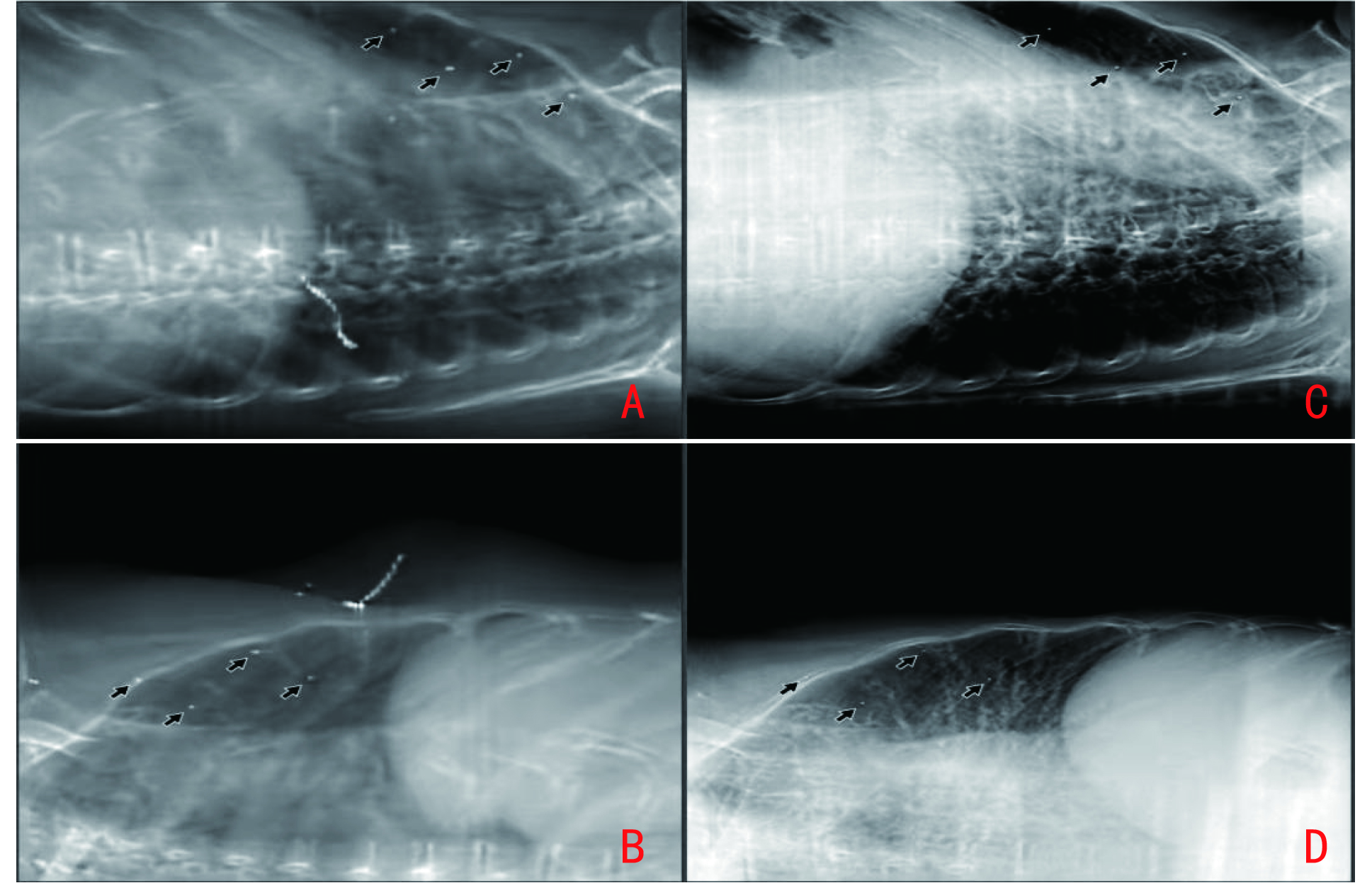
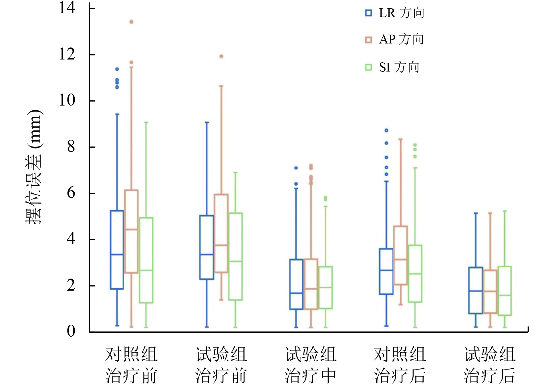
目的 探讨基于图像引导系统的乳腺癌保乳术后行容积旋转调强放疗(volumetric modulated arc therapy, VMAT)患者在投照过程中摆位误差的实时校正及剂量学参数变化。 方法 选取 2020年10月至2021年12月天津医科大学肿瘤医院收治的20例保乳术后行VMAT患者,随机分为对照组10例和试验组10例,放疗时行图像引导,对误差数据进行统计学分析,将摆位误差引入治疗计划重新计算,比较两组剂量学差异。 结果 对照组和试验组在左右(LR)、腹背(AP)、头脚(SI)方向的摆位误差校正前分别为(3.58±2.35)mm和(3.51±2.08)mm、(4.44±3.62)mm 和(4.23±2.17)mm、(2.85±2.36)mm和(2.99±1.90)mm。对照组在治疗后摆位误差分别为(2.64±1.62)、(3.15±1.50)、(2.49±1.70)mm;试验组在治疗中与治疗后摆位误差分别为(2.07±1.65)mm与 (1.85±1.22)mm、(2.29±1.93) mm与(1.78±1.26)mm、(1.98±1.49)mm与(1.67±1.27) mm。LR、AP、SI方向摆位误差≤3 mm时试验组占比多于对照组,两组比较差异均具有统计学意义(χ2=21.07、60.76、33.63,均P<0.01);两组在治疗后摆位误差比较差异亦均具有统计学意义(t=6.36、10.35、5.60,均P<0.05)。试验组在肿瘤区和临床靶区处方剂量的覆盖体积、心脏平均剂量、肺受照剂量均有优势,两组比较差异均具有统计学意义(均P<0.05)。 结论 实时图像引导系统能校正患者投照过程中的摆位误差,治疗中增加一次校正可明显减小分次内摆位误差,并获得更好的剂量学结果。 Abstract:Objective To investigate the setup errors and their dosimetric impacts during volumetric modulated arc therapy (VMAT) for breast cancer after breast-conserving surgery in patients using an image guidance system. Methods Twenty patients who underwent VMAT after breast-conserving surgery at Tianjin Medical University Cancer Institute & Hospital between October 2020 and December 2021 were randomly assigned into the control (n=10) and experimental groups (n=10). The error data were statistically analyzed, and the dosimetric impacts of the positional errors were compared between the two groups by recalculating the treatment plans with the measured setup errors. Results In the control and experimental groups, the setup errors were (3.58±2.35) and (3.51±2.08) mm, (4.44±3.62) and (4.23±2.17) mm, and (2.85±2.36) and (2.99±1.90) mm along the left-right (LR), anterior-posterior (AP), and superior-inferior (SI) directions, respectively. In the control group, the displacement in the LR, AP, and SI directions after irradiation were (2.64±1.62) mm, (3.15±1.50) mm, and (2.49±1.70) mm, respectively. In the experimental group, the displacement in the LR, AP, and SI directions after the first and second arc deliveries were (2.07±1.65) mm and (1.85±1.22) mm, (2.29±1.93) mm and (1.78±1.26) mm, and (1.98±1.49) mm and (1.67±1.27) mm, respectively. The experimental group had more posttreatment positional errors ≤3 mm in the LR, AP and SI directions than the control group, and the difference was statistically significant (χ2=21.07, 60.76, 33.63; P<0.01). The posttreatment displacement exhibited a statistically significant difference between the two groups (t=6.36, 10.35, 5.60; P<0.05). The dosimetric parameters from the recalculated virtual treatment plans showed that the experimental group was significantly superior to the control group in terms of the proportion of gross tumor volume and clinical target volume receiving the prescription dose, the mean dose of the heart, and the dose of the lung (P<0.05). Conclusions The real-time image guidance system can correct setup errors during radiotherapy. Intrafractional correction significantly reduced patient setup errors and obtained better dosimetric results. -
表 1 两组患者治疗后摆位误差比较
方向 对照组* 试验组* χ2 P ≤3 mm >3 mm ≤3 mm >3 mm LR 142(71) 58(29) 180(90) 20(10) 21.07 <0.01 AP 110(55) 90(45) 184(92) 16(8) 60.76 <0.01 SI 130(65) 70(35) 182(91) 18(9) 33.63 <0.01 ()内单位为%;*:均为10例×20次  下载: 导出CSV
表 2 两组患者靶区及重要危及器官剂量学的比较
剂量学指标 对照组 试验组 t P GTV处方剂量体积(%) 88.30±3.80 93.30±3.30 −2.43 0.04 CTV处方剂量体积(%) 85.70±4.00 90.70±2.90 −2.47 0.03 心脏平均剂量(Gy) 5.84±1.85 3.79±1.19 2.29 0.04 患侧肺 平均剂量(Gy) 10.50±2.13 7.78±1.95 2.35 0.04 V5 Gy(%) 46.83±11.12 30.67±13.76 2.24 0.04 V20 Gy(%) 19.29±5.69 13.11±4.92 12.32 <0.01 健侧肺 平均剂量(Gy) 2.90±1.95 2.53±1.86 3.86 0.01 V5 Gy(%) 23.67±10.61 16.32±11.88 1.25 0.24 全肺 平均剂量(Gy) 7.26±1.67 4.97±1.75 2.32 0.04 V5 Gy(%) 35.00±15.35 28.85±19.63 1.14 0.29 V20 Gy(%) 9.83±2.64 6.33±2.25 2.47 0.03
下载: 导出CSV
-
[1] Lei SY, Zheng RS, Zhang SW, et al. Global patterns of breast cancer incidence and mortality: a population-based cancer registry data analysis from 2000 to 2020[J]. Cancer Commun (Lond), 2021, 41(11):1183-1194. doi: 10.1002/cac2.12207 [2] 中国医师协会放射肿瘤治疗医师分会.乳腺癌放射治疗指南(中国医师协会2020版)[J].中华放射肿瘤学杂志,2021,30(4):321-342. doi: 10.3760/cma.j.cn113030-20210107-00010 [3] Speers C, Pierce LJ. Postoperative radiotherapy after breast-conserving surgery for early-stage breast cancer: a review[J]. JAMA Oncol, 2016, 2(8):1075-1082. doi: 10.1001/jamaoncol.2015.5805 [4] Kim H, Lee SB, Nam SJ, et al. Survival of reast-conserving surgery plus radiotherapy versus total mastectomy in early breast cancer[J]. Ann Surg Oncol, 2021, 28(9):5039-5047. doi: 10.1245/s10434-021-09591-x [5] 陈绍芳.锥形束CT引导乳腺癌精确放疗的研究进展[J].中国医学物理学杂志,2019,36(2):163-165. doi: 10.3969/j.issn.1005-202X.2019.02.008 [6] 罗惠煌,童远和,王永斌.早期左侧乳腺癌术后锥形束CT引导下VMAT摆位误差致靶区及危及器官受照剂量变化研究[J].中外医学研究,2020,18(36):66-68. doi: 10.14033/j.cnki.cfmr.2020.36.026 [7] 郭静钰,王伟,孙斌,等.两种千伏级立体平面图像引导放射治疗系统临床一致性研究[J].中国医学装备,2020,17(1):23-27. doi: 10.3969/J.ISSN.1672-8270.2020.01.007 [8] Sarkar B, Munshi A, Ganesh T, et al. Technical note: rotational positional error corrected intrafraction set-up margins in stereotactic radiotherapy: a spatial assessment for coplanar and noncoplanar geometry[J]. Med Phys, 2019, 46(11):4749-4754. doi: 10.1002/mp.13810 [9] 张彦新,方浩,陈冰,等.ExacTrac X-射线图像引导系统在体部肿瘤放疗中的摆位误差和残余误差分析[J].中华放射医学与防护杂志,2019,39(2):95-100. doi: 10.3760/cma.j.issn.0254-5098.2019.02.003 [10] Wennstig AK, Wadsten C, Garmo H, et al. Long-term risk of ischemic heart disease after adjuvant radiotherapy in breast cancer: results from a large population-based cohort[J]. Breast Cancer Res, 2020, 22(1):10. doi: 10.1186/s13058-020-1249-2 [11] González-Sanchis A, Brualla-González L, Fuster-Diana C, et al. Surface-guided radiation therapy for breast cancer: more precise positioning[J]. Clin Transl Oncol, 2021, 23(10):2120-2126. doi: 10.1007/s12094-021-02617-6 [12] Badakhshi H, Gruen A, Sehouli J, et al. The impact of patient compliance with adjuvant radiotherapy: a comprehensive cohort study[J]. Cancer Med, 2013, 2(5):712-717. doi: 10.1002/cam4.114 [13] 董芳芬,戴立言,黄妙云,等.体表铅点标记辅助iSCOUT图像引导定位系统在乳腺癌术后调强放疗应用研究[J].中华放射肿瘤学杂志,2021,30(10):1059-1064. doi: 10.3760/cma.j.cn113030-20210317-00111 [14] Stelczer G, Tatai-Szabó D, Major T, et al. Measurement of dose exposure of image guidance in external beam accelerated partial breast irradiation: evaluation of different techniques and linear accelerators[J]. Phys Med, 2019, 63:70-78. doi: 10.1016/j.ejmp.2019.05.020 [15] Wang W, Yu T, Xu M, et al. Setup error assessment and correction in planar kV image- versus cone beam CT image-guided radiation therapy: a clinical study of early breast cancer treated with external beam partial breast irradiation[J]. Technol Cancer Res Treat, 2019, 18: 1533033819853847. -

 点击查看大图

图(3) / 表(2)
计量
- 文章访问数: 674
- HTML全文浏览量: 18
- PDF下载量: 35
- 被引次数: 0
