
-
摘要:
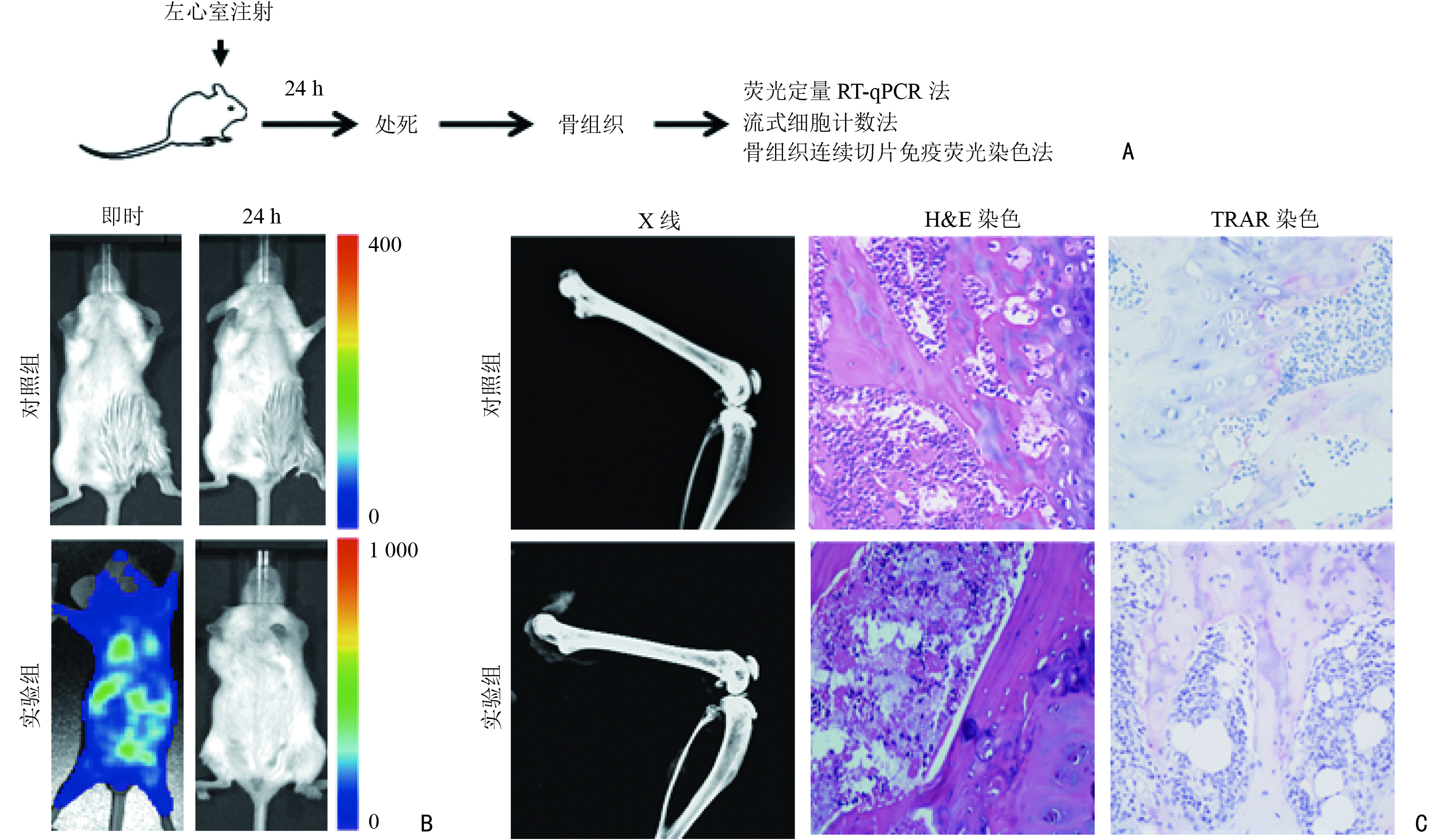
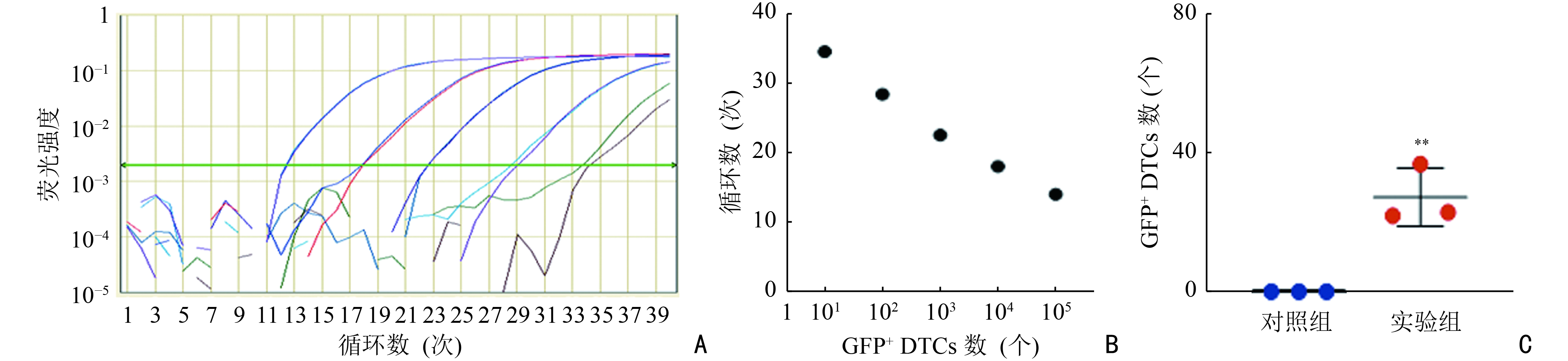
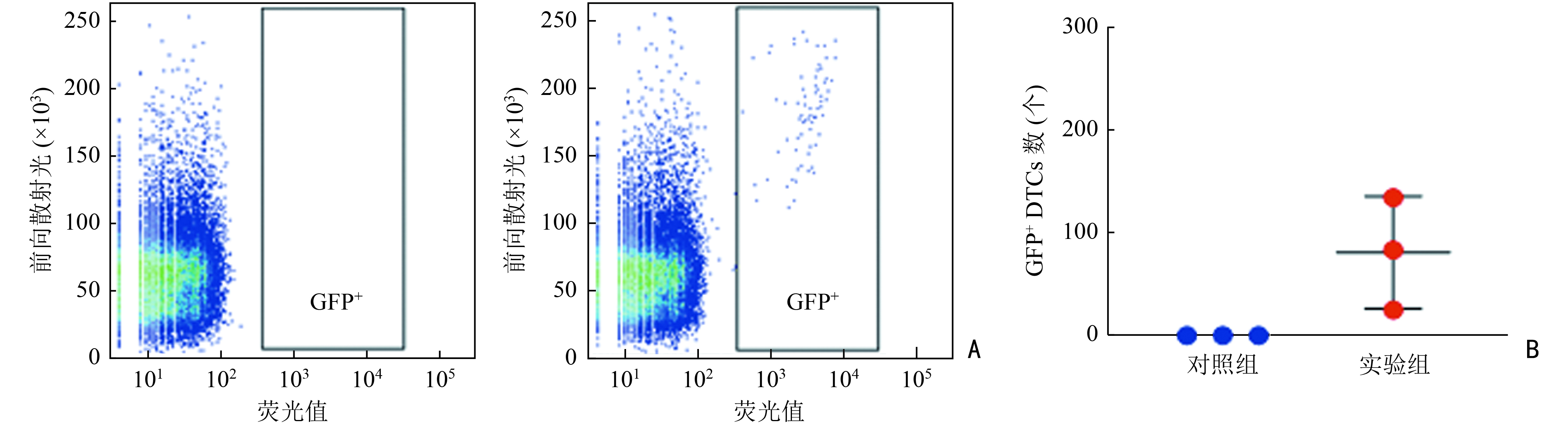
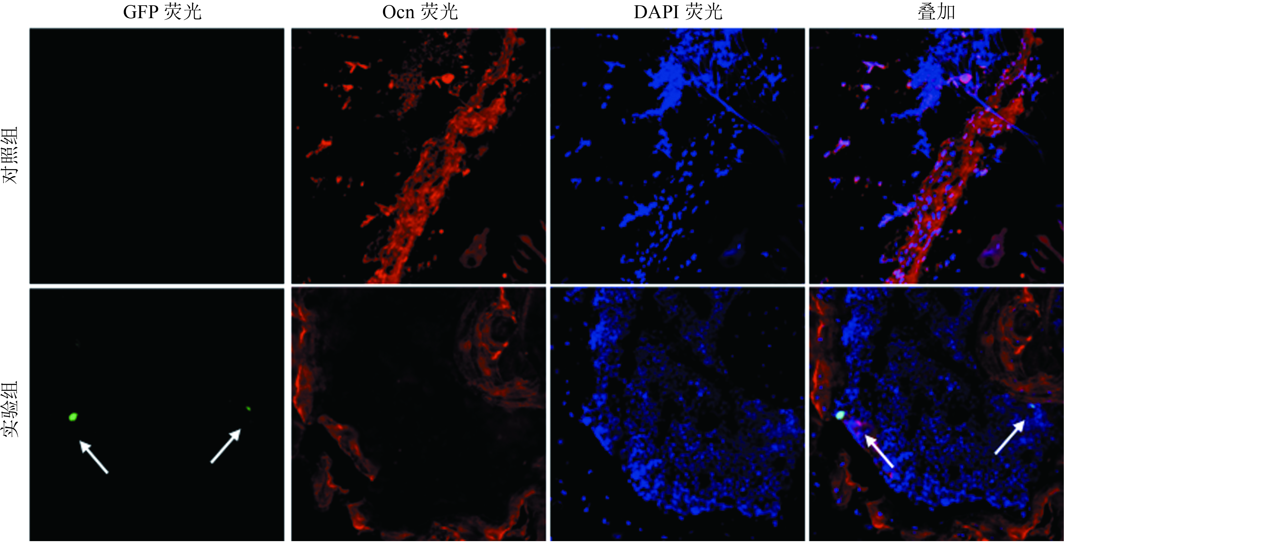
目的 探究小鼠骨髓中播散肿瘤细胞(disseminated tumor cells,DTCs )的检测方法。 方法 采用慢病毒感染的方法构建MDA-MB-231-GFP/Luc乳腺癌细胞系,将MDA-MB-231-GFP/Luc细胞经左心室接种于NOD-SCID小鼠体内,构建骨髓DTCs小鼠模型。采用荧光定量RT-qPCR、流式细胞计数、骨组织连续切片免疫荧光染色三种检测方法对小鼠骨髓DTCs进行定量和组织学定位研究。 结果 荧光定量RT-qPCR法和流式细胞计数法的检测下限分别为22个和25个绿色荧光蛋白阳性(GFP+)细胞。骨组织连续切片免疫荧光染色法虽然不能定量骨髓DTCs数量,但可观察到GFP+ DTCs在骨组织中的分布,定位于成骨细胞或骨基质附近。 结论 三种检测方法联合使用可满足小鼠骨转移研究动物实验中骨髓DTCs的定量和定位研究的需求,可为乳腺癌骨转移和骨髓中休眠癌细胞研究提供方法学支持。 Abstract:Objective To investigate the detection methods of disseminated tumor cells (DTCs) in the bone marrow of mice. Methods The MDA-MB-231-GFP/Luc breast cancer cell line was constructed using lentiviral transfection. MDA-MB-231-GFP/Luc tumor cells were then inoculated into NOD-SCID mice via intracardiac injection in order to construct a mouse model with bone marrow DTCs. Real-time quantitative polymerase chain reaction (RT-qPCR), flow cytometry, and immunofluorescence staining on serial frozen sections of bone tissue were used to quantify DTCs and to observe the histological distribution of DTCs in the bone marrow. Results The detection limits of the RT-qPCR and flow cytometry methods were 22 and 25 GFP+ cells, respectively. Immunofluorescence staining of serial frozen sections of bone tissue did not quantify bone marrow DTCs, but it showed that GFP+ DTCs were localized near osteoblasts or the bone matrix. Conclusions The combination of these three detection methods meets the detection requirements for the quantification and localization of bone marrow DTCs in animal experiments, therefore providing methodological support for research on bone metastasis and dormancy of breast cancer cells in the bone marrow. -
Key words:
- breast cancer /
- bone metastasis /
- bone marrow /
- disseminated tumor cells (DTCs)
-
[1] Coleman RE. Clinical features of metastatic bone disease and risk of skeletal morbidity[J]. Clin Cancer Res, 2006, 12(20 Pt 2): 6243s-6249s. [2] Wang MN, Xia F, Wei YQ, et al. Molecular mechanisms and clinical management of cancer bone metastasis[J]. Bone Res, 2020, 8(1):30. [3] Zhang WJ, Bado IL, Hu JY, et al. The bone microenvironment invigorates metastatic seeds for further dissemination[J]. Cell, 2021, 184(9):2471-2486. doi: 10.1016/j.cell.2021.03.011 [4] Satcher RL, Zhang XHF. Evolving cancer-niche interactions and therapeutic targets during bone metastasis[J]. Nat Rev Cancer, 2022, 22(2):85-101. doi: 10.1038/s41568-021-00406-5 [5] Bado IL, Zhang WJ, Hu JY, et al. The bone microenvironment increases phenotypic plasticity of ER+ breast cancer cells[J]. Dev Cell, 2021, 56(8):1100-1117. doi: 10.1016/j.devcel.2021.03.008 [6] Nobre AR, Risson E, Singh DK, et al. Bone marrow NG2+/Nestin+ mesenchymal stem cells drive DTC dormancy via TGFβ2[J]. Nat Cancer, 2021, 2(3):327-339. doi: 10.1038/s43018-021-00179-8 [7] Li XQ, Zhang R, Lu H, et al. Extracellular vesicle-packaged CDH11 and ITGA5 induce the premetastatic niche for bone colonization of breast cancer cells[J]. Cancer Res, 2022, 82(8):1560-1574. doi: 10.1158/0008-5472.CAN-21-1331 [8] Li XQ, Lu JT, Tan CC, et al. RUNX2 promotes breast cancer bone metastasis by increasing integrin α5-mediated colonization[J]. Cancer Lett, 2016, 380(1):78-86. doi: 10.1016/j.canlet.2016.06.007 [9] Minn AJ, Kang YB, Serganova I, et al. Distinct organ-specific metastatic potential of individual breast cancer cells and primary tumors[J]. J Clin Invest, 2005, 115(1):44-55. doi: 10.1172/JCI22320 [10] Wang H, Yu CJ, Gao X, et al. The osteogenic niche promotes early-stage bone colonization of disseminated breast cancer cells[J]. Cancer Cell, 2015, 27(2):193-210. doi: 10.1016/j.ccell.2014.11.017 [11] Phan TG, Croucher PI. The dormant cancer cell life cycle[J]. Nat Rev Cancer, 2020, 20(7):398-411. doi: 10.1038/s41568-020-0263-0 [12] O'Neill K, Lyons SK, Gallagher WM, et al. Bioluminescent imaging: a critical tool in pre-clinical oncology research[J]. J Pathol, 2010, 220(3):317-327. doi: 10.1002/path.2656 [13] Shao MM, Chan SK, Yu AMC, et al. Keratin expression in breast cancers[J]. Virchows Arch, 2012, 461(3):313-322. doi: 10.1007/s00428-012-1289-9 [14] Mitas M, Mikhitarian K, Walters C, et al. Quantitative real-time RT-PCR detection of breast cancer micrometastasis using a multigene marker panel[J]. Int J Cancer, 2001, 93(2):162-171. doi: 10.1002/ijc.1312 [15] Chen L, Yi XF, Guo P, et al. The role of bone marrow-derived cells in the origin of liver cancer revealed by single-cell sequencing[J]. Cancer Biol Med, 2020, 17(1):142-153. -

 下载:
下载:
 点击查看大图

图(5)
计量
- 文章访问数: 79
- HTML全文浏览量: 8
- PDF下载量: 21
- 被引次数: 0
