
Expression and Clinical Significance of Paxillin and Caspase-3 in Breast Invasive Ductal Carcinoma
-
摘要:
目的 探讨桩蛋白(Paxillin)和天冬氨酸特异性半胱氨酸蛋白酶-3(Caspase-3)在乳腺浸润性导管癌(IDC)中的表达及其与乳腺浸润性导管癌(IDC)患者临床病理因素的关系。 方法 采用免疫组化检测术中切除的原发性乳腺浸润性导管癌(IDC)和距癌灶约5 cm处正常乳腺组织中Paxillin和Caspase-3的表达,统计学分析两者间及其与乳腺浸润性导管癌(IDC)病理特征的关系。 结果 Paxillin在IDC组的表达高于正常乳腺组织组,差异具有统计学意义(P < 0.05);Caspase-3在IDC组的表达低于正常乳腺组织组,差异具有统计学意义(P < 0.05)。在IDC组中,Paxillin的表达与Her-2状态相关,2种蛋白的表达与乳腺IDC的淋巴结转移及组织学分级相关,2种蛋白在IDC中的表达呈负相关(r=-0.32,P=0.013)。 结论 Paxillin和Caspase-3的的异常表达在乳腺浸润性导管癌(IDC)的发生、发展中可能起着重要的作用,且与乳腺浸润性导管癌(IDC)的浸润转移密切相关。 -
关键词:
- 乳腺肿瘤 /
- 桩蛋白 /
- 天冬氨酸特异性半胱氨酸蛋白酶-3
Abstract:Objective To explore the expression and clinical significance of paxillin and caspase-3 in breast invasive ductal carcinoma (IDC). Methods The paxillin and caspase-3 expression in IDC tissue and normal mammary specimens were detected by immunohistochemistry. Then, the correlation between the expression of these two proteins and the clinicopathologic characteristics of breast IDC was analyzed. Results The paxillin expression was significantly higher in IDC tissue than in normal mammary tissue (P < 0.05), whereas the caspase-3 expression was significantly lower in IDC tissue than in normal mammary tissue (P < 0.05). In IDC, the paxillin expression was correlated with the Her-2 level, whereas the paxillin and caspase-3 expression was closely correlated with the lymph-node metastasis and histological grades (P < 0.05). Moreover, negative correlations were observed between the paxillin and caspase-3 expression (r = -0.32; P = 0.013). Conclusion The abnormal expression of paxillin and caspase-3 plays important roles roles in the occurrence and development of IDC and correlates with the invasion and metastasis of breast IDC. -
Key words:
- Breast tumor /
- Paxillin /
- Caspase-3
-

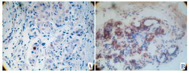
图 2 Caspase-3在乳腺组织中的表达(SP×200)
A:乳腺癌组织;B:正常乳腺组织
Figure 2. Staining of caspase-3(SP×200)
表 1 Paxillin和Caspase-3蛋白表达与乳腺IDC临床病理参数的关系 例(%)
Table 1. The relationship of paxillin and caspase-3 expression with clinicopathologic parameters of breast IDC

表 2 Paxillin和Caspase-3蛋白表达在乳腺IDC中的关系 例
Table 2. The relationship between the expression of paxillin and caspase-3 protein in breast IDC

-
[1] Sattler M, Pisick E, Morrison PT, et al. Role of the cytoskeletal protein paxillin in oncogenesis[J]. Crit Rev Oncol, 2000, 11(1): 63-76. [2] Yang XF, Xin Y, Mao LL. Clinicopathological significance of pten and caspase-3 expressions in breast cancer[J]. Chin Med Sci J, 2008, 23(2) : 95-102. doi: 10.1016/S1001-9294(09)60019-5 [3] McGregor BA, Antoni MH. Psychological intervention and health outcomes among women treated for breast cancer: A review of stress pathways and biological mediators[J]. Brain Behav Immun, 2009, 23(2): 159-166. doi: 10.1016/j.bbi.2008.08.002 [4] Deakin NO, Turner CE. Distinct roles for paxillin and Hic-5 in regulating breast cancer cell morphology, invasion, and metastasis [J]. Mol Biol Cell, 2011, 22(3): 327-341. doi: 10.1091/mbc.e10-09-0790 [5] Livasy CA, Moore D, Cance WG, et al. Focal adhesion kinase overexpression in endometrial neoplasia[J]. Appl Immunohistochem Mol Morphol 2004, 12(4): 342-345. doi: 10.1097/00129039-200412000-00009 [6] 李海刚, 谢德荣, 黎洪浩, 等. 肝细胞癌Paxillin和VEGF的表达及其临床意义[J]. 中国肿瘤临床, 2004, 31(7): 412-413. http://www.cjco.cn/cn/article/doi/ [7] 孙兴华, 王亚红, 马彬, 等. Paxillin和HER-2在乳腺癌中的表达及意义[J]. 辽宁医学院学报, 2009, 30(3): 203-206. doi: 10.3969/j.issn.1674-0424.2009.03.005 [8] Sen A, O'Malley K, Wang Z, et al. Paxillin regulates androgenand epidermal growth factor-induced MAPK signaling and cell proliferation in prostate cancer cells[J]. J Biol Chem, 2010, 285(37): 28787-28795. doi: 10.1074/jbc.M110.134064 [9] Chen T, Wong YS, Zheng W, et al. Caspase and p53-dependent apoptosis in breast carcinoma cells induced by asynthetic selenadiazole derivative[J]. Chem Biol Interact, 2009, 180(1): 54-60. doi: 10.1016/j.cbi.2008.12.010 [10] 应荣彪, 冯俊, 李建军, 等. 胃癌中survivin和caspase-3的表达及其临床意义[J]. 中国癌症杂志, 2010, 20(1): 17-21. doi: 10.3969/j.issn.1007-3639.2010.01.004 [11] 王强, 杨志雄, 廖思海. Fas、FasL和Caspase-3在肺癌中的表达及其临床意义[J]. 现代肿瘤医学, 2009, 17(2): 248-251. doi: 10.3969/j.issn.1672-4992.2009.02.020 [12] Deakin NO, Turner CE. Paxillin comes of age[J]. J Cell Sci, 2008, 121(Pt 15): 2435-2444. http://pubmedcentralcanada.ca/articlerender.cgi?accid=pmc2522309 [13] Phipps LE, Hino S, Muschel RJ. Targeting cell spreading: a method of sensitizing metastatic tumor cells to TRAIL-induced apoptosis [J]. Mol Cancer Res, 2011, 9(3): 249-258. doi: 10.1158/1541-7786.MCR-11-0021 [14] Short SM, Yoder BJ, Tarr SM, et al. The expression of the cytoskeletal focal adhesion protein paxillin in breast cancer correlates with HER2 overexpression and may help predict response to chemotherapy: a retrospective immunohistochemical study[J]. Breast J, 2007, 13(2): 130-139. doi: 10.1111/j.1524-4741.2007.00389.x -

 下载:
下载:
 点击查看大图

图(2) / 表(2)
计量
- 文章访问数: 30
- HTML全文浏览量: 4
- PDF下载量: 0
- 被引次数: 0
