
The Effects of PXD101 on Proliferation and Apoptosis of Human Breast Cell Line MCF-7 and Its Mechanism
-
摘要:
目的 探讨组蛋白去乙酰化酶抑制剂PXD101对人乳腺癌细胞MCF-7增殖、细胞周期及凋亡的影响及分子机制研究。 方法 应用不同浓度PXD101处理培养的乳腺癌细胞株MCF-7, 通过赛唑蓝比色(MTT)法和平板克隆形成实验检测药物对细胞增殖的影响; HoechSt33342荧光染色法观察细胞形态变化; 流式细胞仪PI染色法检测细胞周期变化以及AnnexinV-FITC/PI双染法检测细胞凋亡情况; Westen blot检测p21、CyclinB1、PARP、Bcl-2以及Bax的蛋白表达。 结果 PXD101以剂量时间依赖性抑制MCF-7细胞的增殖; 荧光显微镜观察发现细胞核碎裂, 出现凋亡小体; 0、0.1、1、10μmol/L PXD101作用24 h后, G2/M期细胞比例增加, 分别为(12.66±1.55)%、(20.63±1.32)%、(23.20±1.82)%、(32.19±2.37)%(P < 0.05), 凋亡细胞也增加(P < 0.05);p21表达增多, CyclinBl表达减少, PARP剪切明显增加, Bcl-2表达减少, Bax表达增加。 结论 PXD101在体外条件下能够明显抑制乳腺癌MCF-7细胞的增殖, 诱导细胞周期阻滞及凋亡, 并呈剂量依赖性。 Abstract:Objective This work aims to investigate the effect of PXDIOI, a novel potent histone deacetylase inhibitor, on the cell proliferation, cycle arrest and apoptosis of human breast cancer cell line MCF-7 and to preliminarily explore its molecular mechanism. Methods MCF-7 cells were cultured in RPMI 1640 medium supplemented with 10%fetal bovin serum and were treated with PXDIOI at varying concentrations.The methyl thiazolyl tetrazolium(MTT) assay and clonogenic assay were used to measure cell proliferation. Morphological changes of cells were observed by fluorescent microscope after staining by Hoechst33342.Flow cytometer was used to analyze the cell cycle arrest rates(PI staining) and the cell apoptotic rates(AnnexinV-FITC/PI double-staining).The protein expressions of p21, CyclinB1, PARP, Bcl-2 and Bax were detected by Western blot. Results PXD101 was used to inhibit the proliferation of the MCF-7 cell line in a dose and time-dependent manner.Fluorescence microscope showed there were nuclear fragmentation and apoptosis bodies in the cells.Flow cytometric analysis indicated that PXD101 induced MCF-7 cells in G 2/M phase were significantly increased.After MCF-7 cells exposed to different concentrations of PXD101, i.e., 0, 0.1, 1 and 10μmol/L, for 24 h, the ratio of G2/ M-phase cells was(12.66±1.55)%, (20.63±1.32)%, (23.20±1.82)%and(32.19±2.37)%respectively(P < 0.05).The rates of apoptotic cells were also significantly increased, compared with the control group(P < 0.05).PXDIOI could up-regulate the protein expression of p21 and down-regulate the expression of CyclinBl.The cleavage of PARP and the expression of pro-apoptosis protein Bax were increased while the anti-apoptosis protein Bcl-2 was decreased. Conclusion PXD101 in vitro can significantly inhibit the proliferation and can induce cell cycle arrest and apoptosis on human breast cancer MCF-7 cell line in a dose-dependent manner.PXD101 may become a new anti-tumor drug for human breast cancer. -
Key words:
- Breast neoplasm /
- PXD101 /
- Cell cycle /
- Apoptosis
-

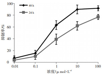
图 1 不同浓度PXD101作用于MCF-7细胞24h和48h后的抑制率曲线
Figure 1. Inhibition ratio of PXD101 on MCF-7 cells at varying concen-trations and in different treatment durations (24h, 48h)

图 2 PXD101作用于MCF-7细胞平板克隆的形成
Figure 2. Clonogenic assay of PXD101 on MCF-7 cells
A: 0 μmol/L; B: 0.1 μmol/L; C: 1 μmol/L; D: 10 μmol/L

图 3 PXD101作用于MCF-7细胞后的周期变化
A:0 μmol/L;B:10 μmol/L 红箭头示G2/M期,绿箭头示subG1峰
Figure 3. Effect of PXD101 on cell cycle distribution of MCF-7. The green arrow shows the subG1 peak

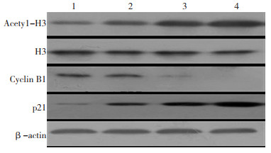
图 4 PXD101作用于MCF-7细胞后Acetyl-H3、H3、CyclinB1及p21蛋白的表达
1:0 μmol/L;2:0.1 μmol/L;3:1 μmol/L;4:10 μmol/L
Figure 4. Effects of PXD101 on the protein expression of Acetyl-H3, H3, Cyclin B1, and p21 in MCF-7 cells

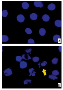
图 5 PXD101作用于MCF-7细胞后细胞形态变化
A:0 μmol/L;B:10 μmol/L;箭头示核碎裂
Figure 5. Morphological changes of MCF-7 cells after PXD101 treatment

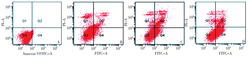
图 6 PXD101作用于MCF-7细胞后的细胞凋亡情况
Figure 6. Apoptosis of MCF-7 cells after PXD101 treatment
A: 0 μmol/L; B: 0.1 μmol/L; C: 1 μmol/L; D: 10 μmol/L

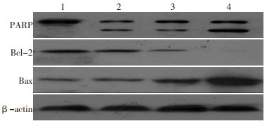
图 7 PXD101作用于MCF-7细胞后PARP、Bcl-2及Bax蛋白的表达
1:0 μmol/L;2:0.1 μmol/L;3:1 μmol/L;4:10 μmol/L
Figure 7. Effects of PXD101 on the protein expression of PARP, Bcl-2 and Bax in MCF-7 cells
表 1 PXD101处理MCF-7细胞24h后细胞周期百分比 (n=3,x±s,%)
Table 1. Cell-cycle distribution of MCF-7 cells at 24h after PXD101 treatment

-
[1] Lane AA, Chabner BA. Histone deacetylase inhibitors in cancer therapy[J]. J Clin Oncol, 2009, 27(32): 5459-5468. doi: 10.1200/JCO.2009.22.1291 [2] Gimsing P. Belmostat: a new broad acting antineoplastic histone deacetylase inhibitor[J]. Expert Opin Investig Drugs, 2009, 18(4): 501-508. doi: 10.1517/13543780902852560 [3] Marks PA, Xu WS. Histone deacetylase inhibitors: Potential in cancer therapy[J]. J Cell Biochem, 2009, 107(4): 600-608. doi: 10.1002/jcb.22185 [4] Singh J, Murata K, Itahana Y, et al. Constitutive expression of the Id-1 promoter in human metastatic breast cancer cells is linked with the loss of NF-1/Rb/HDAC-1 transcription repressor complex[J]. Oncogene, 2002, 21(12): 1812-1822. doi: 10.1038/sj.onc.1205252 [5] Krusehe CA, Wulfing P, Kersting C, et al. Histone deacetylase-1 and -3 protein expression in human breast cancer: a tissue mi-croarray analysis[J]. Breast Cancer Res Treat, 2005, 90(1): 15-23. doi: 10.1007/s10549-004-1668-2 [6] Zhou Q, Dalgard CL, Wynder C, et al. Histone deacetylase inhibitors SAHA and sodium butyrate block G1-to-S cell cycle progression in neurosphere formation by adult subventricular cells[J]. BMC Neurosci, 2011, 12: 50. doi: 10.1186/1471-2202-12-50 [7] Györffy B, Lanczky A, Eklund AC, et al. An online survival analysis tool to rapidly assess the effect of 22, 277 genes on breast cancer prognosis using microarray data of 1, 809 patients[J]. Breast Cancer Res Treat, 2010, 123(3): 725-731. doi: 10.1007/s10549-009-0674-9 [8] Qian X, Ara G, Mills E, et al. Activity of the histone deacetylase inhibitor belinostat(PXD101) in preclinical models of prostate cancer[J]. Int J Cancer, 2008, 122(6): 1400-1410. doi: 10.1002/ijc.23243 [9] Ma BB, Sung F, Tao Q, et al. The preclinical activity of the histone deacetylase inhibitor PXD101(belinostat) in hepatocellular carcinoma cell lines[J]. Ivest New Drugs, 2010, 28(2): 107-114. doi: 10.1007/s10637-009-9219-7 [10] Bi G, Jiang G. The molecular mechanism of HDAC Inhibitors in anticancer effects[J]. Cell Mol Immunol, 2006, 3(4): 285-290. http://www.cnki.com.cn/Article/CJFDTotal-CMIT200604006.htm [11] Tait JF. Imaging of apoptosis[J]. J Nucl Med, 2008, 49(10): 1573-1576. doi: 10.2967/jnumed.108.052803 [12] HwangJJ, Kim YS, Kim MJ, et al. Histone deacetylase inhibitor potentiates anticancer effect of docetaxel via modulation of Bcl-2 family proteins and tubulin in hormone refractory prostate cancer cells[J]. J Urol, 2010, 184(6): 2557-2564. doi: 10.1016/j.juro.2010.07.035 -

 下载:
下载:


 点击查看大图
计量
- 文章访问数: 120
- HTML全文浏览量: 15
- PDF下载量: 0
- 被引次数: 0
