
Expression and Significance of FoxOl and Ki-67 in Esophageal Squamous Cell Carcinoma
-
摘要:
目的 探讨Foxol和Ki-67蛋白在食管鳞状细胞癌组织及食管正常鳞状上皮组织中的表达及其临床意义。 方法 采用免疫组织化学SP法, 检测食管鳞状细胞癌(36例)组织及食管正常鳞状上皮(12例)组织中Foxo1和Ki-67蛋白的表达情况。 结果 Foxol蛋白在食管鳞状细胞癌及食管正常鳞状上皮组织中的阳性表达率分别为75.00%、16.67%, 而Ki-67的阳性表达率分别为83.33%、0。Foxol和Ki-67在食管鳞状细胞癌中的阳性表达率均明显高于食管正常鳞状上皮(P < 0.01)。Foxol蛋白的表达随肿瘤分化程度增高而显著增高(P < O.01), 而Ki-67的表达随肿瘤分化程度增高而降低(P=0.107)。Foxol和Ki-67蛋白的表达与其他临床病理特征无明显相关性(P > 0.05)。 结论 在食管鳞状细胞癌中, Foxol及Ki-67蛋白表达明显高于食管正常鳞状上皮组织, 且随肿瘤分化程度的增高Foxol的表达显著升高, 而Ki-67的表达则显著降低。提示Foxol和Ki-67在食管鳞状细胞癌肿瘤细胞分化过程中发挥着不同作用, 其作用机制不同。 Abstract:Objective To determine the expression and clinical significance of the FoxO1 and Ki-67 proteins in esophageal squamous cell carcinoma(ESCC) and normal esophageal epithelial tissue. Methods Streptavidin-peroxidase(SP) staining was used to detect the expression of FoxO1 and Ki-67 in 36 ESCC samples and 12 samples of normal esophageal epithelium. Results The positive expression rate of FoxOl was 75.00%in ESCC and 16.67%in the normal esophageal epithelium tissue.The positive expression rate of Ki-67 was 83.33%in ESCC and 0%in the normal esophageal epithelium tissue.The positive expression rates of FoxO1 and Ki-67 were significantly higher in ESCC than in normal esophageal tissue(P < 0.01).Among the ESCC cases, the rates of positive FoxO1 and Ki-67 expression were related to the degree of ESCC differentiation.The rate of positive FoxO1 expression increased as the degree of pathological differentiation increased(P < 0.01);however, the rate of positive Ki-67 expression decreased as the degree of pathologic ESCC differentiation increased(P < 0.01).No significant correlation was observed among other clinicopathologic features. Conclusion The positive expression rates of FoxOl and Ki-67 are significantly higher in ESCC than in normal esophageal epithelial tissue.As the degree of pathologic ESCC differentiation increases, the rate of positive FoxOl expression increases, whereas that of Ki-67 decreases. FoxO1 and Ki-67 play different roles in ESCC differentiation, and the mechanism of action is different. -
Key words:
- FoxO1 /
- Ki-67 /
- Esophageal squamous carcinoma /
- Immunohistochemistry
-

图 1 Foxo1在正常食管鳞状上皮组织中的表达(SP ×400)
Figure 1. Foxo1 protein expression in normal esophageal epithelial tissue (SP ×400)

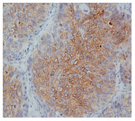
图 2 Foxo1在食管鳞癌组织中的表达(SP ×400)
Figure 2. Foxo1 protein expression in esophageal squamous carcinoma tissue(SP ×400)

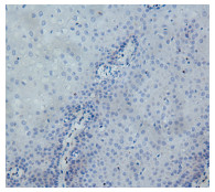
图 3 Ki-67在正常食管鳞状上皮组织中的表达(SP ×400)
Figure 3. Ki-67 protein expression in normal esophageal epithelium tissue(SP ×400)

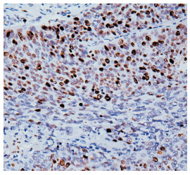
图 4 Ki-67在食管鳞癌组织中高表达(SP ×400)
Figure 4. Ki-67 protein expression in esophageal squamous carcinoma tissue(SP ×400)
表 1 正常食管鳞状上皮和食管鳞癌组织中Foxo1、Ki-67蛋白的表达
Table 1. Foxo1 and Ki-67 protein expression in normal esophageal epithelium and esophageal squamous carcinoma tissue

表 2 Foxo1、Ki-67蛋白的表达与临床病理特征的关系
Table 2. Correlations among Foxo1 and Ki-67 protein expression and the clinicopathologic features of esophageal squamous carcinoma tissue

表 3 食管鳞癌中Foxo1与Ki-67蛋白表达的相关性
Table 3. Correlations between Foxo1 and Ki-67 protein expression in esophageal squamous carcinoma

-
[1] Kaestner KH, Knochel W, Martnez DE. Unified nomenclature for the winged heli x / f orkhead transcription factors[J]. Genes Dev, 2000, 14(2): 142-146. doi: 10.1101/gad.14.2.142 [2] Carroll JS, Liu XS, Brodsky AS, et al. Chromosome-wide mapping of estrogen receptor binding reveals long-range regulation requiring the forkhead protein FoxAI[J]. Cell, 2005, 122(1): 33-43. doi: 10.1016/j.cell.2005.05.008 [3] Fontenot JD, Rasmussen JP, Williams LM, et al. Regulatory T cell lineage specification by the forkhead transcdption factor foxp3[J]. Immunity, 2005, 22(3): 329-341. doi: 10.1016/j.immuni.2005.01.016 [4] 颜亚晖, 钱燕春. 乳腺癌组织中FOX01蛋白表达与细胞增殖和细胞凋亡的相关性[J]. 中国组织化学与细胞化学杂志, 2010, 4(19): 380-384. https://www.cnki.com.cn/Article/CJFDTOTAL-GGZZ201004014.htm [5] Guttilla IK, White BA. Coordinate regulation of FOXO1 by miR-27a, miR-96, and miR-182 in breast cancer cells[J]. J Biol Chem, 2009, 284(35): 23204-23216. doi: 10.1074/jbc.M109.031427 [6] 周英姿, 张友元, 桂律, 等. FOXO1A及FLASH在胃癌组织中的表达及其意义[J]. 肿瘤防治研究, 2010, 6(37): 675-678. https://www.cnki.com.cn/Article/CJFDTOTAL-ZLFY201006022.htm [7] AndersonmJ, Viarsc S, Czekay S, et al. Cloning and characterization of drree human forkhead genes that comprise an FKHR-like gene subfamily[J]. Genomics, 1998, 47(2): 187-199. doi: 10.1006/geno.1997.5122 [8] Duchrow M, Gerdes J, Schluter G, et al. The proliferation-associated Ki-67 protein: definition in molecular terms[J]. Cell Prolif, 1994, 27(5): 235-242. doi: 10.1111/j.1365-2184.1994.tb01421.x [9] 陶玉梅, 陶仪声, 马莉. 食管鳞状细胞癌中MCM2蛋白与Ki-67的表达及对比研究[J]. 临床与实验病理学杂志, 2011, 27(4): 382-384. doi: 10.3969/j.issn.1001-7399.2011.04.010 [10] 郝曙光, 冯笑山, 董彩虹, 等. 食管鳞癌及其癌前病变组织P63与Ki-67蛋白共表达临床意义的探讨[J]. 中华肿瘤防治杂志, 2010, 20 (17): 1646-1661. https://www.cnki.com.cn/Article/CJFDTOTAL-QLZL201020014.htm [11] Verdolini R, Amerio P, Goteri G, et al. Cutaneous carcinomas and preinvasive neoplastic lesions. Role of MMP-2 and MMP-9 metalloproteinases in neoplastic invasion and their relationship with proliferative activity and p53 expression[J]. J Cutan Pathol, 2001, 28 (3): 120-126. doi: 10.1034/j.1600-0560.2001.028003120.x -

 下载:
下载:

 点击查看大图
计量
- 文章访问数: 13
- HTML全文浏览量: 1
- PDF下载量: 0
- 被引次数: 0
