
-
摘要:
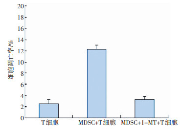
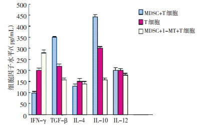
目的 检测乳腺癌患者肿瘤原位组织的一群髓系来源抑制细胞(MDSCs)中吲哚胺2,3双加氧酶(indoleamine-2,3-dioxygenase,IDO)的表达情况,探讨IDO对MDSCs介导T淋巴细胞免疫抑制作用的影响。 方法 收集30例乳腺癌患者的肿瘤组织和外周血及30例健康供者外周血,将肿瘤组织制成单细胞悬液,采用免疫磁珠技术分选肿瘤单细胞悬液中CD33+ MDSCs和健康供者外周血中的CD33+细胞,应用Western blot和PCR方法检测MDSCs中IDO的表达情况。将肿瘤组织来源MDSCs和异体T淋巴细胞按照1:1比例混合培养3天,在加用和不加IDO特异性抑制剂1-MT条件下,利用Annexin-V凋亡试剂盒检测各组T淋巴细胞凋亡率,利用ELISA法检测各组T淋巴细胞分泌的细胞因子量。 结果 Western blot和PCR检测发现MDSCs中IDO过表达。T细胞单独培养时凋亡率为(2.40±0.66)%,MDSCs和T细胞共孵育组中T细胞凋亡率为(12.30±0.80)%,比T细胞单独培养时显著升高(P < 0.05),在共孵育过程中加用1-MT组的T细胞凋亡率为(3.30±0.58)%,与不加1-MT组比较差异具有统计学意义(P < 0.05)。细胞因子检测的结果发现MDSCs促进T淋巴细胞TGF-β、IL-10的释放,抑制IFN-γ的分泌,而对IL-4和IL-12的分泌影响并不明显,而加用1-MT后MDSCs和T淋巴细胞共孵育组中TGF-β、IL-10的分泌水平与未加1-MT组相比显著降低,IFN-γ的分泌显著增加(P < 0.05)。 结论 在乳腺癌患者中,原位肿瘤组织来源的MDSCs对T细胞具有明显的免疫抑制作用;IDO在此群细胞中有过表达,MDSCs发挥免疫抑制作用与IDO密切相关。 -
关键词:
- 髓系来源抑制细胞 /
- 乳腺癌 /
- 吲哚胺2,3-双加氧酶 /
- T细胞
Abstract:Objective This study aimed to explore the secretion of indoleamine-2, 3-dioxygenase (IDO) in myeloid-derived suppressor cells (MDSCs) and its role in immunosuppression and to analyze the relevant impact of MDSCs on T cell proliferation and cytokine secretion. Methods Peripheral blood samples were obtained from 30 breast cancer patients and 30 healthy volunteers from Tianjin Medical University Cancer Institute and Hospital. Breast cancer samples were also acquired from the patients. T cells from the peripheral blood of healthy volunteers and MDSCs from the primary focus of the tumor were separated through a magnetic cell sorting system. IDO expression was determined using Western blot and PCR separately. MDSC-induced T cell apoptosis was detected by flow cytometry. The role of IDO in MDSC immunosuppression was investigated using 1-MT. Cytokine secretion was determined by ELISA. Results Up-regulated IDO expression was found in MDSCs. T cell apoptosis in the group with T cell culture alone, the group with co-culture of MDSCs and T cell, and that with co-culture of MDSCs, T cell, and 1-MT was (2.90 ± 0.66) %, (12.30 ± 0.80) %, and (5.90 ± 0.58) %, respectively. There were significant differences in the T cell apoptosis rate between the group with T cell culture alone and co-culture of MDSCs and T cell. The tumor-derived MDSCs could promote TGF-β and IL-10 secretion and could inhibit IFN-γ secretion dramatically. However, the differences in the secretion of IL-4 and IL-12 were not statistically significant. After incubation with 1-MT, the differences in apoptosis rate between the T cell-alone culture group and the incubation group were not significant. Conclusion IDO expression is upregulated in MDSCs from the primary site of breast cancer. The upregulation of IDO expression may be an important mechanism for the immunosuppression of MDSCs. -
Key words:
- MDSCs /
- Breast cancer /
- Indoleamine 2, 3-dioxygenase (IDO) /
- T cells
-

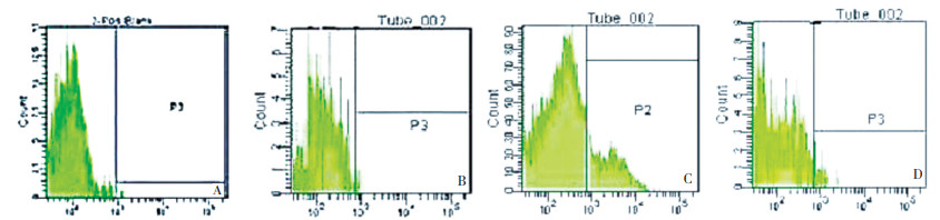
图 1 利用磁珠分选后进行流式检测乳腺癌患者肿瘤单细胞悬液中Lin1-CD45+CD33+CD13+CD14-CD15-所占比例
A:对照组; B:正常人外周血MDSC所占比例; C:乳腺癌患者外周血MDSC所占比例; D:乳腺癌患者肿瘤单细胞悬液中MDSC所占比例
Figure 1. Percentage of MDSC in breast cancer after magnetic cell sorting

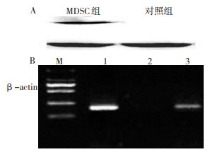
图 2 Western blot(A)和PCR(B)检测MDSC上IDO的表达
M:Marker;1:β-actin;2:对照组IDO;3:实验组IDO
Figure 2. Measurement of IDO expression by MDSC through Western blot and PCR

图 3 MDSCs诱导T细胞凋亡
A:对照组;B:T淋巴细胞单独培养组;C:MDSC+T组;D:MDSC+T+1-MT组
Figure 3. MDSC-induced apoptosis of T cells
-
[1] Srivastava MK, Sinha P, clements VK, et al. Myeloid-derived suppressor cells inhibit T-cell activation by dePleting cystine and cysteine[J]. Cancer Res, 2010, 70(1): 68-77. doi: 10.1158/0008-5472.CAN-09-2587 [2] Hanson EM, Clements VK, Sinha P, et al. Myeloid-derived suPPressor cells down-regulate L-selectin exPression on CD4 + and CD8+ T cells[J]. J Immvnol, 2009, 183(2): 937-944. doi: 10.4049/jimmunol.0804253 [3] Delano MJ, ScumPia PO, Weinstein JS, et al. MyD88-dePendent exPansion of an immature GR-1(+)CD11b(+)PoPulation induces T cell suPPression and Th2 Polarization in sePsis[J]. J Exp Med, 2007, 204(6): 1463-1474. doi: 10.1084/jem.20062602 [4] Gabrilovich DI, Bronte V, Chen SH, et al. The terminology issue for myeloid-derived suPPressor cells[J]. Cancer Res, 2007, 67(1): 425-426. doi: 10.1158/0008-5472.CAN-06-3037 [5] Kusmartsev S, Cheng F, Yu B, et al. All-trans-retinoic acid eliminates immature myeloid cells from tumor-bearing mice and imProves the effect of vaccination[J]. Cancer Res, 2003, 63(15): 4441-4449. http://www.researchgate.net/profile/Sergei_Kusmartsev/publication/10621821_All-_trans_-Retinoic_Acid_Eliminates_Immature_Myeloid_Cells_from_Tumor-bearing_Mice_and_Improves_the_Effect_of_Vaccination/links/0c96051826f87d104e000000.pdf [6] Makarenkova VP, Bansal V, Matta BM, et al. CD11b+/Gr-1+ myeloid suPPressor cells cause T cell dysfunction after traumatic stress [J]. J Immunol, 2006, 176(4): 2085-2094. doi: 10.4049/jimmunol.176.4.2085 [7] Rodriguez PC, Ochoa AC. T cell dysfunction in cancer: role of myeloid cells and tumor cells regulating amino acid availability and oxidative stress[J]. Semin Cancer Biol, 2006, 16(1): 66-72. doi: 10.1016/j.semcancer.2005.10.001 [8] 沈春, 于津浦, 李慧, 等. 乳腺癌患者髓系来源抑制性细胞的鉴定与免疫抑制作用研究[J]. 中国肿瘤临床, 2009, 36(17): 1010-1015. https://www.cnki.com.cn/Article/CJFDTOTAL-ZGZL200917017.htm [9] Sinha P, Clements VK, Bunt SK, et al. Cross-talk between myeloid-derived suPPressor cells and macroPhages subverts tumor immunity toward a tyPe 2 resPonse[J]. J Immunol, 2007, 179(2): 977-983. doi: 10.4049/jimmunol.179.2.977 [10] Movahedi K, Guilliams M, Van den Bossche J, et al. Identification of discrete tumor-induced myeloid-derived suppressor cell subpopulations with distinct T cell-suppressive activity[J]. Blood, 2008, 111(8): 4233-4244. doi: 10.1182/blood-2007-07-099226 [11] Zhu B, Bando Y, Xiao S, et al. CD11b + Ly-6C(hi) suPPressive monocytes in exPerimental autoimmune encePhalomyelitis[J]. J Immunol, 2007, 179(8): 5228-5237. doi: 10.4049/jimmunol.179.8.5228 -

 下载:
下载:
 点击查看大图

计量
- 文章访问数: 55
- HTML全文浏览量: 1
- PDF下载量: 1
- 被引次数: 0
