
Effects of Hydrazinocurcumin-loaded Nanoparticles on the Proliferation and Apoptosis of Breast Cancer Cells in RAW264.7- 4T1 Coculture Conditions
-
摘要:
目的 探索联氨基姜黄素纳米脂质体(hydrazinocurcumin-loaded nanopaticles,HC-NPs)通过抑制STAT3(signal trans? duction and activators of transcription-3)活化,对RAW264.7-4T1共培养体系中4T1细胞增殖及凋亡的影响。 方法 以乳腺癌细胞—巨噬细胞共培养体系中的乳腺癌细胞4T1为研究对象,分PBS对照组和10%NPs对照组、10%HC-NPs处理组。采用检测细胞形态学变化,台盼蓝染色计数细胞存活率,流式细胞仪检测细胞凋亡率及细胞周期变化,Western blot检测STAT3、p-STAT3、c-MYC、Bcl-2及Bax的蛋白表达水平。 结果 刘氏染色发现经10%HC-NPs处理后,4T1细胞形态变圆,台盼蓝染色计数显示细胞存活率减低,且与对照组有显著性差异(P < 0.05);流式细胞仪检测发现HC-NPs处理组细胞凋亡率为(24.21±2.37)%,细胞周期结果显示HC-NPs组处理的细胞主要阻滞在G2/M期为(63.70±2.53)%,与对照组有显著性差异(P < 0.05);Western blot检测显示,HC-NPs组4T1细胞的p-STAT3、c-MYC表达明显减少,Bcl-2/Bax比值下降,而总STAT3水平无明显变化。 结论 HC-NPs能通过抑制STAT3活化(p-STAT3),影响细胞增殖、凋亡相关基因,抑制共培养体系中乳腺癌细胞4T1的增殖,促进其凋亡。 Abstract:Objective To investigate the effects of hydrazinocurcumin-loaded nanoparticles (HC-NPs) on the proliferation and apoptosis of breast cancer 4T1 cells in coculture conditions through the inhibition of the signal transducer and activator of transcription 3 (STAT3) signal pathway activation. Methods Mouse breast cancer 4T1 cells in coculture conditions were divided into three groups, namely, the PBS control, 10% NP control, and 10% HC-NP treatment groups. Morphological investigation was conducted via Liu staining, and the survival ratio was determined via trypan blue staining. Cell cycle and apoptosis were detected using flow cytometry. Western blot analysis was used to determine the expression of STAT3, phosphorylation STAT3 (pSTAT3), c-MYC, B-cell lymphoma 2 (Bcl-2), and Bcl-2-associated X protein (Bax). Results Liu staining results show that the shape of the HC-NP-treated 4T1 cells was spherical. Moreover, trypan blue staining shows that the survival ratio of the treated cells was significantly decreased compared with that of the control. Flow cytometry results show that the apoptosis rates of the HC-NP treatment groups was 24.21% ± 2.37% (P < 0.05), whereas that of The cells in the G2/M phase increased (63.70% ± 2.53%, P < 0.05). Western blot analysis indicates that p-STAT3, c-MYC, and the ratio of Bcl-2/Bax expression decreased. However, that of total STAT3 showed no significant difference with that of the control. Conclusion HC-NPs can inhibit the proliferation and promote the apoptosis of breast cancer 4T1 cells in coculture conditions, possibly by inhibiting STAT3 activity and affecting the genes related to proliferation and apoptosis. -
Key words:
- Hydrazinocurcumin-loaded nanoparticle /
- STAT3 /
- Coculture /
- Breast cancer /
- Cell proliferation /
- Cell apoptosis
-

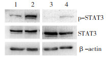
图 2 4T1及RAW264.7细胞共培养前后p-STAT3及STAT3蛋白水平变化
1:4T1;2:4T1与RAW264.7共培养后;3:RAW264.7;4:RAW264.7与4T1共培养后
Figure 2. p-STAT3 and STAT3 expression of 4T1 and RAW264.7 cells before and after co-culture

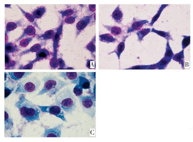
图 3 HC-NPs对共培养体系中4T1细胞形态学影响(×100)
A:PBS对照组;B:10% NPs对照组;C:10% HC-NPs处理组
Figure 3. HC-NPs affects morphology of 4T1 cells in co-culture conditions(×100)

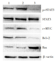
图 4 HC-NPs对共培养体系中4T1细胞中p-STAT3、STAT3、Bax、Bcl-2、c-MYC蛋白水平的影响
1:PBS对照组;2:10% NPs对照组;3:10% HC-NPs处理组
Figure 4. Effects of HC-NPs on p-STAT3, STAT3, Bax, Bcl-2 and c-MYC expression of 4T1 cells in co-culture conditions
表 1 NPs及HC-NPs对共培养体系中4T1细胞凋亡率及细胞周期的影响
% Table 1. Effects of NPs and HC-NPs on apoptosis rate and cell cycle of 4T1 cells in co-culture conditions
(%) 
-
[1] Rathore R, Jain JP, Srivastava A, et al. Simultaneous determination of hydrazinocurcumin and phenol red in samples from rat intestinal permeability studies: HPLC method development and validation[J]. J Pharm Biomed Anal, 2008, 46(2): 374-380. doi: 10.1016/j.jpba.2007.09.019 [2] Zhang B, Wang J, Gao J, et al. Alternatively Activated RAW264.7 Macrophages Enhance Tumor Lymphangiogenesis in Mouse Lung Adenocarcinoma[J]. J Cell Biochem, 2009, 107(1): 134-143. doi: 10.1002/jcb.22110 [3] Luo YP, Zhou H, Krueger J, et al. The role of proto-oncogene Fra-1 in remodeling the tumor microenvironment in support of breast tumor cell invasion and progression[J]. Oncogene. 2010, 29 (5): 662-673. doi: 10.1038/onc.2009.308 [4] Bacman D, Merkel S, Croner R, et al. TGF-beta receptor 2 downregulation in tumour-associated stroma worsens prognosis and high-grade tumours show more tumour-associated macrophages and lower TGF-beta 1 expression in colon carcinoma: a retrospective study[J]. BMC Cancer. 2007, 7: 156. doi: 10.1186/1471-2407-7-156 [5] Herrmann A, Kortylewaki M, Kujawaki M, et al. Targeting STAT3 in the myeloid compartment drastically improves the in vivo antitumor functions of adoptively transferred T cells[J]. Cancer Res, 2010, 70(19): 7455-7464. doi: 10.1158/0008-5472.CAN-10-0736 [6] Dhillon N, Aggarwal BB, Newman RA, et al. Phase Ⅱ trial of curcumin in patients with advanced pancreatic cancer[J]. Clin Cancer Res, 2008, 14(14): 4491-4499. doi: 10.1158/1078-0432.CCR-08-0024 [7] Mackenzie G G, Queisser N, Wolfson M L, et al. Curcumin induces cell-arrest and apoptosis in association with the inhibition of constitutively active NF-kappaB and STAT3 pathways in Hodgkin's lymphoma cells[J]. Int J Cancer, 2008, 123(1): 56-65. doi: 10.1002/ijc.23477 [8] Xu D, Dwyer J, Li H, et al. Ets2 maintains hTERT gene expression and breast cancer cell proliferation by interacting with c-Myc[J]. J Biol Chem, 2008, 283(35): 23567-23580. doi: 10.1074/jbc.M800790200 [9] Fan J, Li R, Zhang R, et al. Effect of Bcl-2 and Bax on survival of side population cells from hepatocellular carcinoma cells[J]. World J Gastroenterol, 2007, 13(45): 6053-6059. doi: 10.3748/wjg.v13.45.6053 [10] Karmakar S, Banik NL, Ray SK. Curcumin suppressed anti-apoptotic signals and activated cysteine proteases for apoptosis in human malignant glioblastoma U87MG cells[J]. Neurochem Res, 2007, 32(12): 2103-2113. doi: 10.1007/s11064-007-9376-z [11] Oltval ZN, Milliaman CL, Korsmeyer SJ. Bcl-2 heterodimerizes in vivo with a conserved homolog, bax, thah acceletates progamed cell death[J]. Cell, 1993, 74(4): 609-619. doi: 10.1016/0092-8674(93)90509-O -

 下载:
下载:
 点击查看大图
计量
- 文章访问数: 33
- HTML全文浏览量: 1
- PDF下载量: 0
- 被引次数: 0
