
Expression of Dihydrofolate Reductase Gene and Its Clinical Significance in Ovarian Cancer
-
摘要:
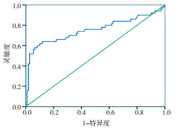
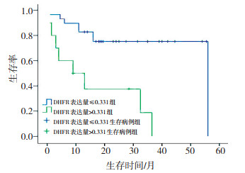
目的 探讨卵巢癌组织中二氢叶酸还原酶基因(dinhydrofolate reductase,DHFR)的表达及其临床意义。 方法 采用实时荧光定量PCR法检测80例卵巢癌组织、50例良性卵巢组织及30例正常卵巢组织中的DHFR mRNA表达情况,分析其与卵巢癌临床病理及多药耐药的相关性。 结果 1)DHFR mRNA在正常卵巢组织、良性卵巢肿瘤和卵巢癌组织中表达量分别为0.584± 0.234、0.895±0.783和0.197±0.412,经比较差异有统计学意义(P < 0.001,P=0.027,P < 0.001)。2)卵巢癌组织中DHFR mRNA表达量与患者临床分期及病理分级差异无统计学意义(P>0.05),但在上皮性癌中浆液性癌组织中DHFR mRNA表达量则明显高于黏液性和内膜样癌,经比较差异有统计学意义(P < 0.001)。3)卵巢癌组织中DHFR mRNA表达量与患者的腹水量、淋巴结转移和远处器官转移无明显相关(P>0.05),但大网膜转移者肿瘤组织中DHFR mRNA表达量则低于无转移者(P=0.044)。4)在卵巢癌患者中治疗后缓解者组织中DHFR mRNA表达量低于肿瘤进展者(P < 0.001);而铂类药物敏感型患者肿瘤组织中DHFR mRNA表达量0.130±0.103也低于铂类药物耐药者0.341±0.701(P=0.011)。5)DHFR mRNA表达量判断卵巢肿瘤性质的ROC曲线Youden指数最大值所对应的数值为0.331,DHFR mRNA表达量>0.331的患者中位生存时间为16.4个月,而≤0.331的患者中位生存时间为44.5个月,差异有统计学意义(P < 0.001),且Cox模型多因素分析亦显示DHFR mRNA表达量为影响预后的独立因素(P=0.018)。 结论 DHFR mRNA在正常和良性卵巢组织中表达量高于卵巢癌组织,提示DHFR参与正常组织细胞生理代谢,而在卵巢癌中DHFR mRNA在铂类耐药组织中表达较高,在铂类敏感组织表达低,提示DHFR与卵巢癌多药耐药相关,可能为判断卵巢癌多药耐药潜在标志之一。 Abstract:Objective The current study aims to investigate the dihydrofolate reductase (DHFR) mRNA expression in ovarian cancer as well as elucidate the relationship between the clinical pathologic parameters and the DHFR mRNA expression in ovarian cancer. Method DHFR expression was detected in 80 cases of ovarian carcinoma, 50 benign ovarian cases and 30 normal controls by real-time fluorescent quantitative polymerase chain reaction. Results The DHFR mRNA expression in ovarian carcinoma, benign ovarian tumor, and normal ovarian tissues was 0.584±0.234, 0.895±0.783, and 0.197±0.412, respectively, with statistically significant differences (P < 0.000, P = 0.027, P < 0.001). No relationship was found between the DHFR mRNA expression and stage or pathological grade of ovarian cancer. But DHFR mRNA expression in serous carcinoma was significantly higher than that in mucinous and endometrioid carcinoma (P = 0.000). DHFR mRNA expression in ovarian cancer was not related with the amount of ascites, lymph node metastasis, or distant metastasis (P > 0.05), but the DHFR mRNA expression in patients with omentum metastasis was lower than that in patients without omentum metastasis, with a statistically significant difference (P = 0.044). DHFR mRNA expression in patients with CR for treatment was lower than that of patients with SD or PD or PR (P < 0.001). The expression of DHFR mRNA in cisplatin-sensitive patients was lower than that in cisplatin-multidrug resistant cases[(0.130 ± 0.103) vs. (0.341 ± 0.701), P = 0.011]. The Youden index of the ROC curve for DHFR mRNA expression was 0.331. The median survival period of patients with DHFR mRNA expression lower than 0.331 was 16.4 months, but the median survival time of patients with DHFR mRNA expression higher than 0.331 was 44.5 months, with a statistically significant difference (P < 0.001). COX multivariate analysis showed that DHFR mRNA expression was a dependent prognostic factor (P = 0.018). Conclusion DHFR plays a role in physiological metabolism in normal cells. A relationship exists between upregulated DHFR mRNA expression and cisplatin-multidrug resistance in ovarian cancer. DHFR mRNA expression could be used as a marker to determine cisplatin-multidrug resistance in ovarian cancer. -

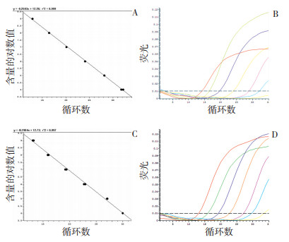
图 1 实时荧光定量PCR测量GAPDH基因及DHFR基因表达量的标准曲线
A、B:GAPDH基因表达量的标准曲线;C、D:DHFR基因表达量的标准曲线
Figure 1. Standard curves of GAPDH and DHFR gene measured by RT-qPCR

图 2 80例卵巢癌组织中DHFR基因表达量的ROC曲线
Figure 2. ROC curve of DHFR gene expression in 80 case of ovarian tissues

图 3 卵巢癌组织中DHFR表达阳性和阴性患者Kaplan-Meier生存曲线
Figure 3. Survival curve of ovarian cancer patients with or without DHFR expression
表 1 实时定量PCR检测各种卵巢组织中DHFR mRNA表达量
x±s Table 1. DHFR transcript expression in various ovarian tissues measured by RT-qPCR

表 2 卵巢癌组织中DHFR mRNA表达量与其临床病理因素的关系
x±s Table 2. The relationship between DHFR mRNA expression and clinicopathologic factors of ovarian cancer

表 3 卵巢癌组织DHFR mRNA表达量与其转移关系
x±s Table 3. The relationship of DHFR mRNA expression in ovarian cancer tissues with metastasis

表 4 卵巢癌组织DHFR mRNA含量与化疗疗效和耐药的相关性
x±s Table 4. The correlation among DHFR mRNA expression, chemotherapy efficacy and drug resistance of ovarian cancer

表 5 Cox比例风险模型分析影响卵巢癌预后的因素
Table 5. Prognostic factors for ovarian cancer analyzed by Cox proportion risk model

-
[1] Askari BS, Krajinovic M. Dihydrofolate reductase gene variations in susceptibility to disease and treatment outcomes[J]. Curr Genomics, 2010, 11(8): 578-583. doi: 10.2174/138920210793360925 [2] Levy AS, Sather HN, Steinherz PG, et al. Reduced folate carrier and dihydrofolate reductase expression in acute lymphocytic leukemia may predict outcome: a Children's Cancer Group Study[J]. J Pediatr Hematol Oncol, 2003, 25(9): 688-695. doi: 10.1097/00043426-200309000-00004 [3] Li L, Luan Y, Li X, et al. Demonstration of differential gene expression between sensitive and resistant ovarian tumor cells by fluorescence differential display-PCR analysis[J]. Oncol Rep, 2005, 13(5): 793-799. [4] Al-Shakfa F, Dulucq S, Brukner I, et al. DNA variants in region for noncoding interfering transcript of dihydrofolate reductase gene and outcome in childhood acute lymphoblastic leukemia[J]. Clin Cancer Res, 2009, 15(22): 6931-6938. doi: 10.1158/1078-0432.CCR-09-0641 [5] Matsuo K, Lin YG, Roman LD, Overcoming platinum resistance in ovarian carcinoma[J]. Expert Opin Investig Drugs, 2010, 19(11): 1339-1354. doi: 10.1517/13543784.2010.515585 [6] Webber S, Bartlett CA, Boritzki TJ, et al. AG337, a novel lipophilic thymidylate synthase inhibitor: in vitro and in vivo preclinical studies[J]. Cancer Chemother Pharmacol, 1996, 37(6): 509-517. doi: 10.1007/s002800050422 [7] Matakidou A, El Galta R, Rudd MF, et al. Prognostic significance of folate metabolism polymorphisms for lung cancer[J]. Br J Cancer, 2007, 97(2): 247-252. doi: 10.1038/sj.bjc.6603830 [8] Serra M, Reverter Branchat G, Maurici D, et al. Analysis of dihydrofolate reductase and reduced folate carrier genstatusin relation to methotrexate resistance in osteosarcoma cells[J]. Ann Oncol, 2004, 15(1): 151-160. doi: 10.1093/annonc/mdh004 [9] Banerjee D, Mayer Kuckuk P, Capiaux G, et al. Noveaspects of resistance to drug stargeted t o dihydrofolate reductase and thymidylate synthase[J]. Biochim Biophys Acta, 2002, 1587(2-3): 164-173. doi: 10.1016/S0925-4439(02)00079-0 [10] 王镜岩, 朱圣庚, 徐长法, 主编. 生物膜的组成与结构, 生物化学(上册) [M]. 第3版. 北京: 高等教育出版社, 2006: 613-614. [11] Wu MF, Hsiao YM, Huang CF, et al. Genetic determinants of pemetrexed responsiveness and nonresponsiveness in non-small cell lung cancer cells[J]. J Thorac Oncol, 2010, 5(8): 1143-1151. doi: 10.1097/JTO.0b013e3181e0b954 [12] Liu X, Yu X, Guo Z, et al. Cotransduction of Human mdr-1 and Dihydrofolate Reductase Genes into Murine Hematopoietic Cells[J]. Zhongguo Shi Yan Xue Ye Xue Za Zhi. 2000, 8(2): 110-113. [13] 于明东, 李书忠. 实时荧光定量PCR法检测人骨肉瘤耐MTX细胞系中RFC、DHFR、GST-π的mRNA表达[J]. 中国矫形外科杂志, 2009, 17(11): 858-861. https://www.cnki.com.cn/Article/CJFDTOTAL-ZJXS200911031.htm [14] 黄瀚, 李兵, 欧阳林旗, 等. 叶酸受体及二氢叶酸还原酶与人乳腺癌细胞MCF-7/ADR多要耐药关系[J]. 中国药理学通报, 2011, 27(1): 99-103. https://www.cnki.com.cn/Article/CJFDTOTAL-YAOL201101028.htm [15] Scionti I, Michelacci F, Pasello M, et al. Clinical impact to the methotrexate resistance associated genes CMYC and dihydrofolate reductase(DHFR) in high grade osteosarcoma[J]. Ann Onco, 2008, 19 (8): 1500-1508. doi: 10.1093/annonc/mdn148 [16] 张文卿, 许佐良, 于杰, 等. 人突变DHFR基因高效逆转录病毒载体及其在小鼠造血细胞的表达[J]. 中国肿瘤生物治疗杂志, 1997, 4(12)∶ 85-89. https://www.cnki.com.cn/Article/CJFDTOTAL-ZLSW199702002.htm [17] Hagner N, Joerger M. Cancer chemotherapy: targeting folic acid synthesis[J]. Cancer Manag Res, 2010, 2: 293-301. http://pdfs.semanticscholar.org/b5fd/ae4511e0ccca684672f3b08bc8e1a5037e12.pdf [18] Schmeler KM, Gershenson DM. Low-grade serous ovarian cancer: a unique disease[J]. Curr Oncol Rep, 2008, 10(6): 519-523. doi: 10.1007/s11912-008-0078-8 -

 下载:
下载:
 点击查看大图
计量
- 文章访问数: 15
- HTML全文浏览量: 1
- PDF下载量: 0
- 被引次数: 0
