
Relationship of Oct-4 and Sox-2 Expression in Colon Carcinoma with Recurrence and Metastasis
-
摘要:
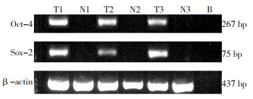
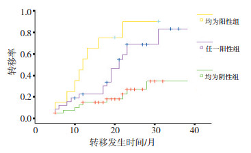
目的 探讨结肠癌组织中Oct-4、Sox-2表达情况及其对术后复发转移的预测作用。 方法 采用免疫组织化学方法检测手术切除的80例结肠癌术后标本中Oct-4、Sox-2表达情况,并且对两指标的表达同肿瘤分化程度、分期和术后复发转移关系进行分析。采用RT-PCR检测20例冰冻肿瘤组织及癌旁组织中Sox-2、Oct-4表达情况。 结果 80例患者中35例出现复发转移,Sox-2、Oct-4在转移组中表达率分别为48.57%(17/35)、51.43%(18/35),在非转移组中表达率分别为17.78%(8/45)、13.33%(6/ 45),差异有统计学意义(P=0.001)。原发病灶分化程度、T分期和N分期同Oct-4和Sox-2表达无显著性差异。均为阳性表达者其转移发生率高,生存分析显示不同表达状态转移出现时间差异有统计学意义(P=0.001)。RT-PCR分析显示20例肿瘤组织中Sox-2、Oct-4阳性表达率分别为40%(8/20)、45%(9/20),明显高于癌旁正常组织5%(1/20)。 结论 肿瘤组织中Sox-2、Oct-4表达同结肠癌术后转移相关,两者联合检测有助于评估结肠癌术后复发转移的可能。 Abstract:Objective The current work aims to investigate the expression of Sox-2 and Oct-4 in colon carcinoma and its relationship with relapse and metastasis. Methods The expression of Oct-4 and Sox-2 was detected in 80 cases of colon cancer tissue by immunohistochemical assay. The correlation of the Oct-4 and Sox-2 expression with histological differentiation, T stage, N stage, and metastasis was analyzed. The level of Oct-4 and Sox-2 expression in the cancer and normal paraneoplastic tissues was evaluated using RT-PCR. Results Metastasis was observed in 35 of all the 80 cases studied. The positive rate of Sox-2 and Oct-4 in the metastasis group was 48.57% (17/35) and 51.43% (18/35), respectively, whereas it was 17.78% (8/45) and 13.33% (6/45) in the non-metastasis group. Significant differences were observed between the two groups (P = 0.001). The expression of Oct-4 and Sox-2 was not associated with differentiation, T stage, or N stage of the tumor. The group with positive expression of Oct-4 and Sox-2 showed high metastatic rate. The survival analysis showed that the time of metastasis was significantly different in the groups with various expressions (P = 0.001). The positive expression rate of Sox-2 and Oct-4 detected by RT-PCR in colonic carcinoma tissues was 40% (8/20) and 45% (9/20) respectively, higher than that in normal tissues. Conclusion The expression of Sox-2 and Oct-4 in tumor tissue can predict postoperative relapse and metastasis of colon carcinoma. -
Key words:
- Colon carcinoma /
- Oct-4 /
- Sox-2 /
- Metastasis /
- Recurrence
-

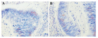
图 1 Oct-4、Sox-2在结肠癌组织中表达(S-P×400)
A:Oct-4阳性;B:Sox-2阳性
Figure 1. Expression of Oct-4 and Sox-2 in colon cancer

图 2 RT-PCR检测Oct-4、Sox-2在结肠癌组织及癌旁组织中的表达
T:肿瘤组织;N:癌旁组织;B:空白对照
Figure 2. Expression of Oct-4 and Sox-2 in colon cancer and normal paraneoplastic tissues

图 3 Oct-4、Sox-2阳性表达和阴性表达患者转移出现时间比较(以转移为截点)
Figure 3. Comparison of metastasis between patients with Oct-4 and Sox-2 expression and those without Oct-4 and Sox-2 expression(P=0.001)(metastasis was censored)
表 1 引物序列
Table 1. Primer sequences

表 2 组织中Sox-2、Oct-4表达同临床特征间关系
Table 2. Relationship of Sox-2 and Oct-4 expression with clinical characteristics

-
[1] 张楠, 连鹏, 时永香, 等. Oct-4/Sox-2协同调控下游基因表达的分子机制[J]. 生物物理学报, 2007, 23(6): 420-426. doi: 10.3321/j.issn:1000-6737.2007.06.002 [2] 王海威, 王家东. Oct-4蛋白在甲状腺肿瘤中的表达及意义[J]. J Clin Otorhinolaryngol Head Neck Surg(China), 2010, 24(15): 682-685. https://www.cnki.com.cn/Article/CJFDTOTAL-LCEH201015006.htm [3] Ben-Porath I, Thomson MW, Carey VJ, et al. An embryonic stem cell-like gene expresson signature in poorly differentiated aggressive human tumors[J]. Nat Genet, 2008, 40(5): 499-507. doi: 10.1038/ng.127 [4] 曹婧, 樊青霞, 索振河. 靛玉红对膀胱癌ScaBer细胞株增殖的影响及机制[J]. 山东医药, 2008, 48(14): 61-62. doi: 10.3969/j.issn.1002-266X.2008.14.031 [5] 曹浩哲, 冀静, 郑鹏生. Oct4基因在宫颈癌中的表达及其意义[J]. 西安交通大学学报(医学版), 2010, 31(1): 17-21. https://www.cnki.com.cn/Article/CJFDTOTAL-XAYX201001006.htm [6] 宋娟, 严宁, 张汉东, 等. 口腔鳞癌组织中干细胞转录因子Oct-4的表达及意义[J]. 临床口腔医学杂志, 2010, 26(7): 390-393. doi: 10.3969/j.issn.1003-1634.2010.07.002 [7] 侯轶, 赵晓昆, 蒋宏毅, 等. 膀胱移行细胞癌组织中PSCA和Oct-4表达及其意义[J]. 实用肿瘤杂志, 2010, 25(5): 531-533. https://www.cnki.com.cn/Article/CJFDTOTAL-SYZZ201005010.htm [8] Attasi Y, Mowla SJ, Ziaee SA, et al. Oct-4, an embryonic stem cell marker, is highly expressed in bladder cancer[J]. Int J Cancer, 2007, 120(7): 1598-1602. doi: 10.1002/ijc.22508 [9] Li XL, Eishi YB, Bai YQ, et al. Expression of the SRY-related HMG box protein SOX2 in human gastric carcinoma[J]. Int J Oncol, 2004, 24(2): 257-263. http://www.spandidos-publications.com/ijo/24/2/257/download [10] Sattler HP, Lensch R, Rohde V, et al. Novel amplification unit at chromosome 3q25-q27 in human prostate cancer[J]. Prostate, 2000, 45(3): 207-215. doi: 10.1002/1097-0045(20001101)45:3<207::AID-PROS2>3.0.CO;2-H [11] Jung M, Peterson H, Chavez L, et al. A data integration approach to mapping OCT4 gene regulatory networks operative in embryonic stem cells and embryonal carcinoma cells[J]. PloS One, 2010, 5(5): e10709. doi: 10.1371/journal.pone.0010709 [12] Yamaguchi S, Kurimoto K, Yabuta Y, et al. Conditional knockdown of Nanog induces apoptotic cell death in mouse migrating primordial germ cells[J]. Development, 2009, 136(23): 4011-4020. doi: 10.1242/dev.041160 [13] Park IH, Zhao R, West JA, et al. Reprogramming of human somatic cells to pluripotency with defined factors[J]. Nature, 2008, 451 (7175): 141-146. doi: 10.1038/nature06534 [14] Lengerke C, Fehm T, Kurth R, et al. Expression of the embryonic stem cell marker SOX-2 in early-stage breast carcinoma[J]. BMC cancer, 2011, 11: 42. doi: 10.1186/1471-2407-11-42 [15] Fong H, Hohenstein KA, Donovan PJ. Regulation of self-renewal and pluripotency by Sox2 in human embryonic stem cell[J]. Stem Cells, 2008, 26(8): 1931-1938. doi: 10.1634/stemcells.2007-1002 -

 下载:
下载:
 点击查看大图
计量
- 文章访问数: 15
- HTML全文浏览量: 2
- PDF下载量: 0
- 被引次数: 0
