
-
摘要:
目的 检测人卵巢癌组织中PGRMC1(progesterone receptor membrane component-1)基因表达情况,探讨PGRMC1基因在卵巢癌发生发展过程中的作用。 方法 利用免疫组织化学方法检测78例不同病变卵巢组织蜡块中PGRMC1基因及蛋白表达情况;以TBP(TATA box binding protein)为内源性对照基因,利用实时定量PCR方法检测了10例正常新鲜卵巢组织和30例新鲜卵巢癌组织中PGRMC1基因的mRNA表达水平。 结果 采用免疫染色强度加着色面积的积分标准判断免疫组织化学结果,与正常卵巢(0.20±0.71)及卵巢良性肿瘤组(0.75±1.12)相比,卵巢交界性肿瘤中PGRMC1蛋白表达(3.60±1.14)和卵巢癌(7.30±2.12)显著增高(P < 0.01)。实时定量PCR方法证实卵巢癌组PGRMC1 mRNA表达(3.526±1.386)显著高于良性卵巢病变组(0.936±0.725)(P < 0.01),是良性卵巢病变组织表达的3.51倍。PGRMC1蛋白阳性表达的卵巢组织中,正常卵巢组MVD为(18.6±6.7)条;良性卵巢病变组MVD为(20.9±7.9)条;交界性卵巢肿瘤组MVD为(22.4±4.7)条;卵巢癌组MVD为(28.4±8.1)条,卵巢癌组与正常卵巢组、良性卵巢肿瘤组间差异有统计学意义(P < 0.01)。 结论 PGRMC1高表达可能与卵巢癌的发生、发展有关,并可能促进血管生成。 Abstract:Objective To examine the expression of progesterone receptor membrane component-1 (PGRMC1) gene and explore its effect on the evolution of ovarian cancers. Methods The protein expression of PGRMC1 in various ovarian lesions was detected by immunohistochemistry. TATA box binding protein gene was used as an endogenous control. The mRNA expression of PGRMC1 gene in 10 cases of benign ovarian tumor tissues and 30 cases of epithelial ovarian cancer tissue was quantified by real-time PCR. Results The expression levels of PGRMC1 protein in borderline ovarian tumor (3.60 ± 1.14) and ovarian cancer (7.30 ± 2.12) tissues were significantly higher than in normal ovarian (0.20 ± 0.71) and benign ovarian tumor (0.75 ± 1.12) tissues (all P < 0.01). Real-time PCR confirmed that the mRNA expression level was significantly different between ovarian cancer (3.526 ± 1.386) and benign ovarian tumor (0.936 ± 0.725) tissues (P < 0.01). The microvessel density in ovarian cancer tissues (28.4 ± 8.1) was significantly higher than in normal ovarian (18.6 ± 6.7) and benign ovarian tumor (20.9 ±7.9) tissues (all P < 0.01). Conclusion The expression of PGRMC1 gene in ovarian tumor tissues is significantly elevated, which may play an important role in the occurrence, development, and angiogenesis of ovarian cancer. -
Key words:
- Ovarian tumor /
- PGRMC1 gene /
- MVD
-

图 1 卵巢良性肿瘤组织PGRMC1蛋白的阳性表达(×200)
Figure 1. Expression of PGRMC1 protein in benign ovarian tumor tissues

图 2 卵巢癌组织PGRMC1蛋白的阳性表达(×200)
Figure 2. Expression of PGRMC1 protein in epithelial ovarian cancer tissues

图 3 RT-PCR检测PGRMC1 mRNA在卵巢组织中的表达
M:marker;1:卵巢癌组;2:良性卵巢病变组
Figure 3. RT-PCR examination of PGRMC1 expression in different ovarian tissues

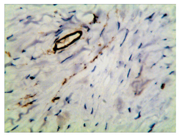
图 4 卵巢癌组织中CD34阳性MVD(×400)
Figure 4. Expression of CD34 in epithelial ovarian cancer tissues

图 5 卵巢纤维瘤中CD34阳性MVD的阳性表达(×400)
Figure 5. Expression of CD34 protein in uterine fibroids tissues
表 1 正常卵巢组织和各种卵巢病变组织中PGRMC1蛋白的表达
Table 1. Protein expression of PGRMC1 in various ovarian lesions

表 2 PGRMC1蛋白表达与卵巢癌病理参数间的关系
例(%) Table 2. Relationship between the protein expression of PGRMC1 and clinicopathologic features of ovarian cancer

-
[1] Hughes AL, Powell DW, Bard M, et al. Dap1/PGRMC1 binds and regulates cytochrome P450 enzymes[J]. Cell Metab. 2007, 45(5): 143-149. http://www.onacademic.com/detail/journal_1000035368249510_9741.html [2] Peluso JJ, Pappalardo A, Losel R, et al. Progesterone membrane receptor component 1 expression in the immature rat ovary and its role in mediating progesterone's antiapoptotic action[J]. Endocrinology. 2006, 147(6): 3133-3140. doi: 10.1210/en.2006-0114 [3] 张鸣号, 彭亮, 曹军. 显微图像分析法与人工计数法在免疫组化结果判读中的应用[J]. 宁夏医科大学学报, 2009, 4(2): 261-262. doi: 10.3969/j.issn.1674-6309.2009.02.057 [4] Peluso JJ, Romak J, Liu X. Progesterone receptor membrane component-1 (PGRMC1) is the mediator of progesterone's antiapoptotic action in spontaneously immortalized granulosa cells as revealed by PGRMC1 small interfering ribonucleic acid treatment and functional analysis of PGRMC1 mutations[J]. Endocrinology, 2008, 149 (3): 534-543. [5] Weidner N, Semple JP, Welch WR, et al. Tumor Angiogenesis and metastasis-correlation in invasive breast carcinoma[J]. N Engl J Med, 1991, 324(1): 1-8. doi: 10.1056/NEJM199101033240101 [6] Skibbens RV. Cell biology of cancer: BRCA1 and sister chromatid pairing reactions[J]? Cell Cycle, 2008, 7: 449-452. doi: 10.4161/cc.7.4.5435 [7] Palmer JE, Sant Cassia IJ, lrwin CJ, et al. Prognostic value of measuerments of angiogenesis in serous carcinoma of the ovary[J]. Int J Gynecol Pathol, 2007, 26(4): 395-403. doi: 10.1097/pgp.0b013e318063bed7 [8] Ace CI, Okulicz WC. Microarray profiling of progesterone-regulated endometrial genes during the rhesus monkey secretory phase[J]. Reprod Biol Endocrinol, 2004, 46(2): 54-56. [9] Schumacher M, Guennoun R, Stein DG, et al. Progesterone: therapeutic opportunities for neuroprotection and myelin repair[J]. Pharmacol Ther, 2007, 116(7): 77-106. [10] Irby RB, Malek RL, Bloom G, et al. Iterative microarray and RNA interference-based interrogation of the SRC-induced invasive phenotype[J]. Cancer Res, 2005, 65(3): 1814-1821. http://cancerres.aacrjournals.org/cgi/reprint/65/5/1814.pdf -

 下载:
下载:


 点击查看大图

计量
- 文章访问数: 19
- HTML全文浏览量: 4
- PDF下载量: 0
- 被引次数: 0
