
-
摘要: 神经纤维瘤是来源于周围神经的良性肿瘤, 可发生于身体任何部位。神经纤维瘤伴多发钙化临床少见, 当其位于四肢软组织时极易误诊, 且临床症状及影像学表现无特征, 术前准确诊断较为困难本文回顾性分析天津医科大学附属肿瘤医院骨与软组织肿瘤科1例经手术切除且病理证实的神经纤维瘤伴多发钙化的临床特点及影像学表现, 同时结合相关文献, 对神经纤维瘤伴钙化病例进行整理分析, 以期初步探讨神经纤维瘤伴多发钙化的临床特征、影像学表现及鉴别诊断要点, 提高对本病的认识。Abstract: Neurofibroma is a benign tumor derived from the peripheral nerve and could occur in any part of the body.Neurofibroma with multiple calcifications is uncommon and is easily misdiagnosed.Accurate preoperative diagnosis for neurofibroma is difficult because of lack of clinical symptoms and imaging characteristics.This study aims to retrospectively analyze the clinical features and imaging performance of one neurofibroma case confirmed via surgery and pathology in the Department of Bone and Soft Tissue Tumor of our hospital. Related studies were reviewed to explore the clinical features, imaging findings, and differential diagnosis of neurofibroma with multiple calcifications and to improve the understanding of this disease.
-
Key words:
- Neurofibroma /
- Calcification /
- Imaging
-

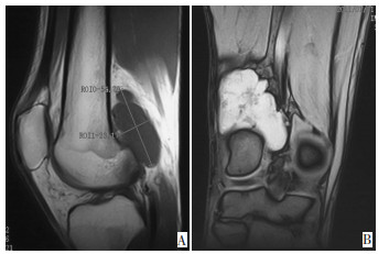
图 1 MRI显示股骨后侧不规则多结节肿物,T1W1呈低信号,T2WI呈不均匀高信号
A:MRI T1W1矢状位;B:MRI T2WI冠状位
Figure 1. MRI shows irregular nodular tumor of the femur posterior: T1W1 at low signal and T2W1 at inhomogeneous high signal

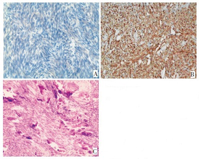
图 2 神经纤维瘤组织病理学特点
:神经纤维瘤细胞梭长杆状,胞浆NSE阳性(IHC×200);B:肿瘤细胞胞浆/胞核弥漫性表达S-100(IHC ×100);C:肿瘤细胞间大量点状、片状钙化(H&E×100)
Figure 2. Histopathological characteristics of neurofibroma
表 1 神经纤维瘤伴钙化影像学特点
Table 1. Imaging characteristics of neurofibroma with calcification

-
[1] Lin J, Martel W. Cross-sectional imaging of peripheral nerve sheath tumors: characteristic signs on CT, MR imaging, and sonography[J]. AJR Am J Roentgenol, 2001, 176(1): 75-82. doi: 10.2214/ajr.176.1.1760075 [2] 余文昌, 王仁法. 巨大少见神经纤维瘤的CT诊断(附3例报告)[J]. 放射学实践, 2000, 15(1): 2. https://www.cnki.com.cn/Article/CJFDTOTAL-FSXS200001038.htm [3] 刘吉华, 房世保, 徐文坚, 主编. 软组织肿瘤成像[M]. 北京: 人民卫生出版社, 2004: 227-239. [4] 王海峰, 方健, 徐军, 等. 小腿神经纤维瘤6次复发后恶变1例[J]. 斛放军医学杂志, 2009, 34(10): 1. https://www.cnki.com.cn/Article/CJFDTOTAL-JFJY200910039.htm [5] Reynolds RM, Browning GG, Nawroz I, et al. Von Recklinghausen's neurofibromatosis: neurofibromatosis type 1[J]. Lancet, 2003, 361(9368): 1552-1554. doi: 10.1016/S0140-6736(03)13166-2 [6] Shibano S, Iguchi T, Nakatani T. A case of scrotal neurofibroma originating from subcutaneous neural tissue[J]. Int J Urol, 2010, 17 (4): 387-388. doi: 10.1111/j.1442-2042.2010.02481.x [7] Gartner L, Pearce CJ, Saifuddin A. The role of the plain radiograph in the characterisation of soft tissue tumours[J]. Skeletal Radiol, 2009, 38(6): 549-558. doi: 10.1007/s00256-008-0513-9 [8] Steenbrugge F. Verstraete K, Poffyn B, et al. Recurrent massive subperiosteal hematoma in a patient with neurofibromatosis[J]. Eur Radiol. 2001, 11(3): 480-483. [9] 韩丹, 王国强, 陆琳. 鼻腔、鼻窦神经纤维瘤的CT表现[J]. 放射学实践, 2003.18(5): 2. https://www.cnki.com.cn/Article/CJFDTOTAL-FSXS200305023.htm [10] Kapoor R. Mittal KP, Jayaram G. Solitary neurofibroma of foot-an unusual case with extensive calcification and ossification[J]. Australas Radiol, 1986, 30(2): 150-152. doi: 10.1111/j.1440-1673.1986.tb02409.x [11] 王兴基, 王平, 吴锋, 等. 多发性尺神经纤维瘤伴钙化一例报告[J]. 中国罕少见病杂志, 1995, 2(2): 2. [12] 徐万鹏, 李佛保, 主编. 骨与软组织肿瘤学[M]. 北京: 人民卫生出版社, 2008: 424-433. [13] 董鹏, 王滨, 常光辉, 等. 盆腔腹膜外间隙神经源性肿瘤的CT诊断[J]. 实用放射学杂志, 2007, 23: 469-474. doi: 10.3969/j.issn.1002-1671.2007.04.012 [14] Rha SE, Byun JY, Jung SE, et al. Neurogenic tumors in the abdomen: tumor types and imaging characteristics[J]. Radiographics, 2003, 23(1): 29-43. doi: 10.1148/rg.231025050 -

 下载:
下载:
 点击查看大图
计量
- 文章访问数: 65
- HTML全文浏览量: 2
- PDF下载量: 1
- 被引次数: 0
