
Whole-body Magnetic Resonance Imaging in Detecting Distant Metastases of Untreated Nasopharyngeal Carcinoma
-
摘要:
目的 探讨磁共振全身成像对鼻咽癌初诊患者远处转移的诊断价值。 方法 2008年11月至2009年8月对福建省肿瘤医院经病理证实的291例鼻咽癌初诊患者在治疗前一周内行全身磁共振成像(Whole Body Magnetic Resonance Imaging,WB-MRI)、X线胸片、肝脏超声和99mTc-MDP核素骨扫描,记录每例患者检出的骨、肺、肝转移灶数目,采用SPSS 15.0统计软件,对几种影像方法各自检出的骨、肺、肝转移灶数目行McNemar's检验,比较检出率的差异。 结果 本组研究共检出骨转移者24例,共发现骨转移灶95个,其中WB-MRI和骨扫描分别检出71个和36个,两者之间差异有统计学意义(P=0.004)。WB-MRI诊断出肺转移6例,而X线胸片诊断出3例,两者间差异无统计学意义(P>0.25);WB-MRI诊断出肝转移6例,而肝脏超声诊断处4例,两者间差异亦无统计学意义(P>0.05)。此外,WB-MRI和超声分别检出肝血管瘤12例和24例(P < 0.01),以及肝囊肿34例和16例(P < 0.005)。 结论 WB-MRI在检测鼻咽癌全身远处转移有较高的应用价值,可作为其M分期的首选影像学检查。 Abstract:Objective To evaluate the clinical value of whole body magnetic resonance imaging (WB - MRI) in detecting distant metastases of initial nasopharyngeal carcinoma (NPC). Methods Examinations of WB-MRI, bone scan, chest X-ray, and liver ultrasound were conducted on 291 consecutive NPC patients within one week before relative treatments. Two experienced radiologists interpreted the WB-MRI. Additional conventional MR or CT was performed on areas with abnormal lesions. Feasibility of metastatic lesions was pathologically confirmed, and all patients were clinically followed up with imaging modalities for at least 12 months. The number of bone, lung, and hepatic metastases detected by WB-MRI were separately compared with those detected by bone scan, chest X-ray, and liver ultrasound. All results were recorded and statistically analyzed by McNemar's test on SPSS (Version 15.0). Results A total of 24 positive patients with bone metastases and 95 lesions of bone metastases were diagnosed by both WB-MRI and bone scan. Among these, 71 lesions were detected through WB-MRI and 36 lesions through bone scan. The difference between the results was significant, with WB-MRI higher than bone scan (P=0.004). Apart from those with bone metastases, six patients with lung metastases were diagnosed by WB-MRI and three patients by chest X-ray. No significant difference was observed (P>0.25). WB-MRI also diagnosed six hepatic metastases patients, whereas liver ultrasound detected only four. Similarly, there was no significant difference between them (P>0.5). Furthermore, 24 patients were diagnosed with hepatic hemangioma through liver ultrasound, and 12 patients were diagnosed by WB-MRI, showing a significant difference between them (P < 0.01). By contrast, 34 patients with hepatic cysts were diagnosed by WB-MRI, and 16 patients were detected by liver ultrasound, showing a significant difference between them (P < 0.005). Conclusion WB-MRI is valuable in detecting distant metastases within the whole body, and it can serve as the first-line imaging technique for the M staging of untreated NPC patients. -

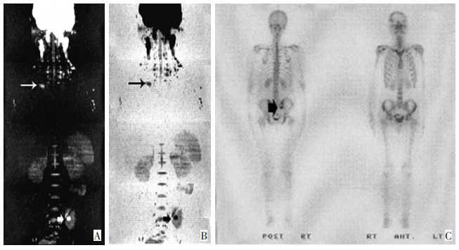
图 1 女性,22岁,鼻咽非角化性未分化型癌
A、B分别为WB-DWI 3D-MIP重建图及WB-DWI 3D-MIP重建黑白反转图,显示右侧锁骨胸骨端(长箭头)及左侧髂骨处转移灶及其形态(小短箭头)。C为核素骨扫描图像:仅显示左侧髂骨异常浓聚(大短箭头)。
Figure 1. Female, 22 years old, non-keratinizing undifferentiated nasopharyngeal carcinoma

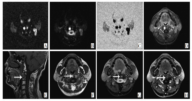
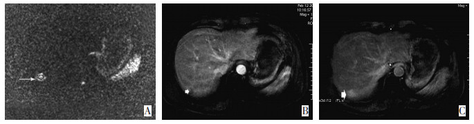
图 2 男性,44岁,鼻咽非角化性未分化型癌
A:DWI图(b值为800s/mm2),显示了右肝第Ⅶ段两枚转移结节呈明显高信号(小短箭头所示);同时可见T12右侧椎弓根转移(长箭头所示)及腹腔、腹膜后间隙多发淋巴结转移灶(大短箭头所示)均呈明显高信号。B:增强后的LAVA图像,显示右肝两枚转移结节的边缘不规则强化(小短箭头所示);T12右侧椎弓根转移灶(长箭头所示)及腹腔、腹膜后间隙淋巴结转移灶(大短箭头所示)亦见不规则明显强化。C:为ADC(Apparent Diffusion Coefficient)图融合同层LAVA图,显示右肝第Ⅶ段两枚转移结节、T12右侧椎弓根转移灶及腹腔、腹膜后淋巴结转移灶明显强化呈高信号,重叠ADC图后有助于ADC值测量,定位更加精确,病灶的境界更加清晰。
Figure 2. Male, 44 years old, non-keratinizing undifferentiated nasopharyngeal carcinoma

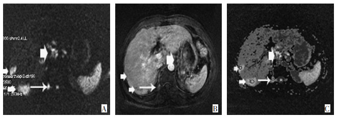
图 3 男性,52岁,鼻咽非角化性未分化型癌
A:b值为800 s/mm2DWI图;、B:b值为0 s/mm2DWI图;C:DWI黑白反转图。短箭头显示:第二颈椎结节状高信号;D:增强后LAVA图像,显示第二颈椎结节状强化(短箭头所示。E、F分别为常规MR检查的矢状位和轴位T1WI-FSE图像,显示第二颈椎及附件低信号(长箭头所示);G:轴位PDWI(fs)图像,显示第二颈椎及附件结节状高信号(长箭头所示);H:为轴位增强T1WIfs+C图像,显示第二颈椎病灶结节状强化(长箭头所示)。
Figure 3. Male, 52 years old, non-keratinizing undif⁃ ferentiated nasopharyngeal carcinoma
-
[1] Vokes EE, Liebowitz DN, Weichselbaum RR, et al. Nasopharyngeal carcinoma. Lancet[J]. 1997, 350(9084): 1087-1091. doi: 10.1016/S0140-6736(97)07269-3 [2] Feng-Yuan Liu, Joseph T. Chang, Hung-Ming Wang, et al. [18F] Fluorodeoxyglucose Positron Emission Tomography Is More Sensitive Than Skeletal Scintigraphy for Detecting Bone Metastasis in Endemic Nasopharyngeal Carcinoma at Initial Staging. Journal of Clinical Oncology. 2006, 24(4): 599-604. doi: 10.1200/JCO.2005.03.8760 [3] 卢秋霞, 卫光宇, 唐溢聪, 等. 148例鼻咽癌肝转移的治疗与预后分析. 实用肿瘤学杂志[J]. 2009, 23(4): 314-318. [4] 邱素芳, 潘建基, 唐明灯, 等. 鼻咽癌343例放射性核素骨显像的临床分析. 肿瘤研究与临床[J]. 2008, 20(5): 331-333. doi: 10.3760/cma.j.issn.1006-9801.2008.05.014 [5] 黄启洪, 李艳华, 林晓, 等. 广东省四会市1995-2004年鼻咽癌生存率分析. 中国肿瘤[J]. 2007, 16(3): 150-152. doi: 10.3969/j.issn.1004-0242.2007.03.003 [6] Takahara T, Imai Y, Yamashita T, et al. Diffusion weighted whole body imaging with background body signal suppression (DWIBS): Technical improvement using free breathing, STIR and high resolusion 3D display. Rad Med[J]. 2004, 22(4): 275-282. http://europepmc.org/abstract/MED/15468951 [7] Xue HD, Li S, Sun HY, et al. Clinical application of body diffusion weithted MR imaging in the diagnosis and preoperative N staging of cervical cancer. Chin J Med Sci[J]. 2008, 23(3): 133-137. doi: 10.1016/S1001-9294(09)60027-4 [8] Shu J, Zhao JN, Zhang XM, et al. The evaluation of LAVA CEMRA angiography in normal peri-pancreatic arteries. J Clin Radiol[J]. 2006, 25(8): 742-745. http://en.cnki.com.cn/Article_en/CJFDTOTAL-LCFS200608019.htm [9] Li Y, Vijayakumar S, Huang F, et al. Reconstrution in image space using basis functions for partially parallel imaging. Magn Reson Imaging[J]. 2008, 26(4): 461-473. doi: 10.1016/j.mri.2007.10.001 [10] Lauenstein TC, Goehde SC, Herborn CU, et al. Whole-body MR imaging : evaluation of patients for matastases. Radiology[J]. 2004, 233 (1): 139-148. doi: 10.1148/radiol.2331030777 -

 下载:
下载:
 点击查看大图
计量
- 文章访问数: 70
- HTML全文浏览量: 2
- PDF下载量: 0
- 被引次数: 0
