
Diagnosis of Lymph Node Metastasis in Lung Cancer by Fine Needle Aspiration Cytology and LunX mRNA Expression
-
摘要:
目的 探讨细针吸取细胞学(fine needle aspiration cytology,FNAC)结合LunX mRNA定量检测在诊断肺癌淋巴结转移中的应用价值。 方法 选择46例肺癌患者进行研究,其中鳞癌22例,腺癌17例,小细胞癌4例,低分化癌不能明确类型3例;同期20例非肺癌患者为对照组。利用细针吸取细胞学穿刺取材,结合细胞学和应用逆转录PCR定量检测穿刺样本LunX mRNA表达水平,确定肺癌患者是否存在淋巴结转移。 结果 肺癌组穿刺标本中LunX mRNA表达的阳性率为84.8%,对照组穿刺标本的阳性率仅为5%,两组阳性率具有显著性差异(χ2=37.16,P < 0.01),并且肺癌转移组与对照组相比较,LunX mRNA平均拷贝数具有显著性差异(Z=-5.807,P < 0.01)。肺癌淋巴结转移患者中LunX mRNA表达的阳性率分别为转移性鳞癌86.4%(19/22)、腺癌70.6%(12/17)、小细胞癌75.0%(3/4)、低分化癌66.7%(2/3),各组间无显著性差异(P=0.482),且各组间LunX mRNA平均拷贝数亦无显著性差异(F=0.377,P=0.770)。 结论 细针吸取细胞学联合LunX mRNA定量检测判断肺癌淋巴结转移是一种微创、快速、准确的手段,有较高的敏感性和特异性。 Abstract:Objective This work investigates the diagnostic significance of fine needle aspiration cytology (FNAC) and LunX mRNA for lymph node metastasis in lung cancer. Methods FNAC was used to examine the lymph nodes of lung cancer patients; quantitative real-time reverse transcription polymerase chain reaction (RT - PCR) was employed to determine the LunX mRNA levels in the biopsy materials. Both these methods could detect whether lymph node metastasis occurred among lung cancer patients. Results In examining the lymph nodes, LunX mRNA was detected in 84.8% of the patients with lung cancer, whereas only in 5 % patients in the control group (χ2 = 37.16, P < 0.01). LunX mRNA Objective: copies in the group with metastatic lung cancer were significantly different with the control group (Z = - 5.807, P < 0.01). Furthermore, the positive rates for LunX mRNA in different histologic types were as follows: 86.3 % (19 / 22) in metastatic squamous cell carcinoma, 70.6 % (12 / 17) in adenocarcinoma, 75 % (3 / 4) in small cell carcinoma, and 66.7 % (2 / 3) in poorly differentiated carcinoma. No significant difference was observed in the statistical data (P = 0.482) and LunX mRNA copy numbers (F = 0.377, P = 0.770) of these groups. Conclusion FNAC combined with the detection of LunX mRNA levels is a minimally invasive, rapid, and accurate diagnostic method for diagnosing lymph node metastasis in lung cancer. -
Key words:
- FNAC /
- LunX /
- RT - PCR /
- Lung cancer /
- Lymph node metastasis
-

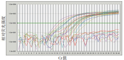
图 1 部分肺癌患者和对照病例LunX基因扩增曲线
Figure 1. Amplification curves observed during LunX assay for lung cancer patients and control

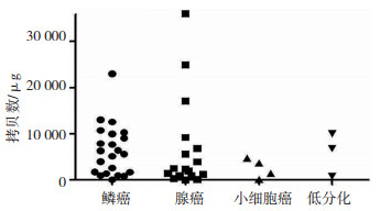
图 2 肺癌淋巴结转移组与对照组穿刺物LunX基因的表达
Figure 2. Expression of LunX genes in lung cancer group with metasta⁃ sized nodes and the control group

图 3 肺癌淋巴结转移组不同组织类型穿刺物LunX的表达
Figure 3. Expression of LunX genes in nodal metastasis of lung cancer of various histological types
表 1 765例淋巴结FNAC结果 例
Table 1. Results of fine needle aspiration cytology in 765 patients

表 2 269例恶性病变FNAC结果 例
Table 2. Fine needle aspiration cytology of 269 patients with malignant neoplasms

表 3 66例患者LunX基因检测结果
Table 3. Expression of LunX mRNA in 66 patients

-
[1] Iwao K, Watanabe T, Fujiwara Y, et al. Isolation of a novel human lung- specific gene, LUNX, a potential molecular marker for detection of micrometastasis in non-small-cell lung cancer[J]. Int J Cancer, 2001, 91(4): 433-437. doi: 10.1002/1097-0215(200002)9999:9999<::AID-IJC1059>3.0.CO;2-B [2] Cheng M, Chen Y, Yu X, et al. Diagnostic utility of LunX mRNA in peripheral blood and pleural fluid in patients with primary non-small cell lung cancer[J]. BMC Cancer, 2008, 8: 156. doi: 10.1186/1471-2407-8-156 [3] 余小琴, 程民, 张艳斌, 等. LunX和CK19基因定量表达在鉴别良恶性胸水中的应用[J]. 中华胸心血管外科杂志, 2007, 23(5), 327-328. doi: 10.3760/cma.j.issn.1001-4497.2007.05.013 [4] 余小琴, 程民, 张艳斌, 等. LunX和CK19基因定量表达在肺癌诊断及疗效观察中的应用研究[J]. 中华肿瘤防治杂志, 2008, 15(4): 255-258. doi: 10.3969/j.issn.1673-5269.2008.04.005 [5] 张洁霞, 于洪波, 何建行. 非小细胞肺癌Lunx mRNA表达临床意义的研究[J]. 中华肿瘤防治杂志, 2009, 16(22): 1774-1777. https://www.cnki.com.cn/Article/CJFDTOTAL-QLZL200922015.htm [6] Mitas M, Hoover L, Silvestri G, et al. Lunx is a superior molecular marker for detection of non-small cell lung cancer in peripheral blood[corrected][J]. J Mol Diagn, 2003, 5(4): 237-242. doi: 10.1016/S1525-1578(10)60480-1 [7] 余辉, 黄秀英, 胡袆, 等. 检测外周血Lunx mRNA表达在诊断非小细胞肺癌微转移中的临床价值[J]. 中国肿瘤临床, 2012, 39(2): 74-76. doi: 10.3969/j.issn.1000-8179.2012.02.004 [8] Lv M, Wu MZ, Zhao YJ, et al. Expression and clinical significance of lung-specific X protein mRNA in bronchial brushing specimens from patients with or without lung cancer[J]. Respirology, 2011, 16(7): 1076-1080. doi: 10.1111/j.1440-1843.2011.02008.x [9] 朱广迎, 刘德林, 王绪, 等. LunX mRNA RT-PCR检测肺癌的微转移[J]. 中国肿瘤临床, 2003, 30(2): 124-127. https://www.cnki.com.cn/Article/CJFDTOTAL-ZGZL200302014.htm [10] Benlloch S, Galbis-Caravajal JM, Alenda C, et al. Expression of molecular markers in mediastinal nodes from resected stage I non-small-cell lung cancer(NSCLC): prognostic impact and potential role as markers of occult micrometastases[J]. Ann Oncol, 2009, 20(1): 91-97. doi: 10.1093/annonc/mdn538 [11] 王潍博, 崔言刚, 姚舒洋. 肺特异性X蛋白角蛋白19及癌胚抗原基因在非小细胞肺癌淋巴结微转移中的比较[J]. 中华肿瘤杂志, 2008, 30 (2): 121-124. -

 下载:
下载:

 点击查看大图
计量
- 文章访问数: 20
- HTML全文浏览量: 1
- PDF下载量: 0
- 被引次数: 0
