
Photodynamic damage of carcinophotorin on breast cancer cells MDA-MB-231 and its mechanism
-
摘要:
目的 探讨癌光啉(PSD-007)在体外对于乳腺癌细胞株MDA-MB-231光动力杀伤效应, 并分析其分子机制。 方法 MTT法检测不同浓度PSD-007(0、2、4、6、8、10μg/mL)作用于MDA-MB-231细胞株后对其增殖的影响; 光学显微镜下观察光动力治疗后细胞形态的变化, 荧光显微镜分析PSD-007作用后细胞的死亡形式。应用RT-PCR和Western blotting技术分析其分子机制。 结果 当PSD007浓度为10μg/mL、光照能量为9.0 J/cm2时, 对乳腺癌细胞的光动力杀伤效应最大, 抑制率为97.01%。光动力治疗处理后的细胞逐渐变圆, 体积变小, 核质比增大, 折光性减弱, 贴壁能力下降, 细胞间隙增大, 直到细胞漂浮死亡。荧光显微镜分析结果显示, 光动力治疗后死亡细胞主要为坏死或晚期的凋亡细胞。RT-PCR和Western blot结果显示相比对照组, 实验组Caspase3、Caspase8、P65亚基和P53表达水平明显上调, 而Bcl-2和Bcl-x表达无明显改变。 结论 PSD-007在体外通过调控Caspase蛋白酶、P53、NF-KB通路对人乳腺癌MDA-MB-231细胞具有光动力杀伤效应, 可能成为未来乳腺癌治疗的一个新方式。 -
关键词:
- 光动力疗法 /
- 乳腺癌 /
- 癌光啉PSD-007 /
- 凋亡
Abstract:Objective his study aims to evaluate the efficacy of carcinophotorin(PSD-007) photosensitization on the apoptosis-induced response in human breast cancer cells and analyze the mechanisms of PSD-007 involved in this process. Methods Methyl thiazolyl tetrazolium(MTT) assay and in situ labeling were performed to examine the effects of the photodynamic therapy(PDT) on the proliferation and apoptosis of the cancer cells MDA-MB-231, respectively.Changes in cellular morphology were assessed using an optical microscope.Real-time polymerase chain reaction and Western blot analysis were conducted to clarify the underlying mechanisms. Results The MTT assay revealed that at a concentration of 10 μg/mL and in combination with 9.0 J/cm2 laser radiation power 24 h after cell culture, PSD-007 markedly inhibited the proliferation of breast tumor cells, with an inhibition rate of 97.01%. "Under a fluorescent microscope, apoptotic cells in the treatment groups with 5 and 10 μg/mL PSD-007-PDT were observed to have dramatically outnumbered the control groups.The dead cells after PSD-007-PDT mainly consisted of necrotic and late apoptotic cells.Caspase-3, caspase-8, P65, and P53 expression was upregulated in the treatment groups compared with the control group, with 5 and 10 μg/ml PSD-007 therapies, whereas no significant alteration in Bcl-2 and Bcl-x was found. Conclusion PDT inhibits the proliferation and induces the apoptosis of cancer cells MDA-MB-231 by upregulating the caspase-3, caspase-8, P53, and NF-KB pathways, indicating a new strategy for treating breast cancer in the future. -
Key words:
- photodynamic therapy /
- breast cancer /
- carcinophotorin/PSD-007 /
- apoptosis
-

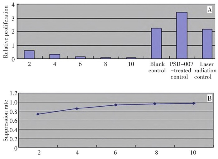
图 1 不同浓度PSD-007对乳腺癌细胞MDA-MB-231增殖
Figure 1. Inhibition rate of PSD-007-PDT with different concentrations on the proliferation of breast cancer cells MDA-MB-231
A: In vitro; B: Proliferation inhibition rate

图 2 荧光显微镜下观察PSD-007-PDT对乳腺癌细胞凋亡的影响
Figure 2. Effects of PSD-007-PDT on the apoptosis of breast cancer cells observed under a fluorescent microscope
A: Group with 5 μ g/mL PSD-007-PDT; B: Group with 10 μ g/mL PSD-007-PDT; C: Blank control; D: Laser irradiation control; E: Control group with PSD-007 treatment

图 3 PSD-007实验组和对照组相对表达量
Figure 3. PSD-007-PDT and the controls
A: Expression of caspase-3, caspase-8, P65, P53, Bcl-2, and Bcl-x mRNA in the groups with 5 and 10 μg/mL PSD-007-PDT and the con⁃ trols; B: Expression of caspase-3, caspase-8, P65, P53, Bcl-2, and Bcl-x proteins in the groups with 5 and 10 μg/mL
表 1 各基因和内参引物
Table 1. Genes and internal control primers

-
[1] Jung NC, Kim HJ, Kang MS, et al. Photodynamic therapy-mediat ed DC immunotherapy is highly effective for the inhibition of estab lished solid tumors[J]. Cancer Lett, 2012, 324(1): 58-65. doi: 10.1016/j.canlet.2012.04.024 [2] Daziano JP, Günther WH, Krieg M, et al. Photochemically generat ed elemental selenium forms conjugates with serum proteins thatare preferentially cytotoxic to leukemia and selected solid tumorcells[J]. Photochem Photobiol, 2012, 88(2): 448-460. doi: 10.1111/j.1751-1097.2012.01078.x [3] Vittar NB, Awruch J, Azizuddin K, et al. Caspase-independent apoptosis, in human MCF-7c3 breast cancer cells, following photodynamic therapy, with a novel water-soluble phthalocyanine[J]. Int J Biochem Cell Biol, 2010, 42(7): 1123-1131. doi: 10.1016/j.biocel.2010.03.019 [4] Livak KJ, Schmittgen TD. Analysis of relative gene expression datausing real-time quantitative PCR and the 2(-Delta Delta C(T))Method[J]. Methods, 2001, 25(4): 402-408. doi: 10.1006/meth.2001.1262 [5] Tirapelli LF, Morgueti M, Cunha Tirapelli DP, et al. Apoptosis inglioma cells treated with PDT[J]. Photomed Laser Surg, 2011, 29(5): 305-309. doi: 10.1089/pho.2009.2649 [6] Kim KH, Park JJ. The effects of photodynamic therapy inupper-gastrointestinal malignant diseases[J]. Gut Liver, 2010, 4 Suppl1: S39-S43. [7] Yang L, Wei Y, Xing D, et al. Increasing the efficiency ofphotodynamic therapy by improved light delivery and oxygensupply using an anticoagulant in a solid tumor model[J]. Lasers Surg Med, 2010, 42(7): 671-679. [8] Weiss A, den Bergh HV, Griffioen AW, et al. Angiogenesis inhibition for the improvement of photodynamic therapy: Therevival of a promising idea[J]. Biochim Biophys Acta, 2012, 1826(1): 53-70. [9] Kessel D, Oleinick NL. Initiation of autophagy by photodynamictherapy[J]. Methods Enzymol, 2009, 453: 1-16. [10] Ji HT, Chien LT, Lin YH, et al. 5-ALA mediated photodynamictherapy induces autophagic cell death via AMP-activated proteinkinase[J]. Mol Cancer, 2010, 9: 91. doi: 10.1186/1476-4598-9-91 [11] Lavi A, Weitman H, Holmes RT, et al. The depth of porphyrin in amembrane and the membrane's physical properties affect thephotosensitizing efficiency[J]. Biophys J, 2002, 82(4): 2101-2110. doi: 10.1016/S0006-3495(02)75557-4 [12] Tang PM, Liu XZ, Zhang DM, et al. Pheophorbide a basedphotodynamic therapy induces apoptosis via mitochondrial-mediatedpathway in human uterine carcinosarcoma[J]. Cancer Biol Ther, 2009, 8(6): 533-539. doi: 10.4161/cbt.8.6.7694 [13] 蔡雄伟, 刘承宜, 丁新民, 等. 血卟啉单甲醚2光动力疗法对Bcl-2的作用[J]. 光电子. 激光, 2005, 16(3): 376-379. https://www.cnki.com.cn/Article/CJFDTOTAL-GDZJ20050300T.htm [14] Hoi SW, Wong HM, Chan JY, et al. Photodynamic Therapy ofPheophorbide a Inhibits the Proliferation of Human BreastTumour via Both Caspase-dependent and-independent Apoptotic Pathways in In Vitro and In Vivo Models[J]. PhytotherRes, 2012, 26(5): 734-742. [15] Weyhenmeyer B, Murphy AC, Prehn JH, et al. Targeting theanti-apoptotic bcl-2 family members for the treatment of cancer[J]. Exp Oncol, 2012, 34(3): 192-199. [16] García-Sáez AJ. The secrets of the Bcl-2 family[J]. Cell DeathDiffer, 2012, 19(11): 1733-1740. [17] Li B, Chu X, Gao M, et al. Apoptotic mechanism of MCF-7 breastcells in vivo and in vitro induced by photodynamic therapy with C-phycocyanin[J]. Acta Biochim Biophys Sin(Shanghai), 2010, 42(1): 80-89. [18] Derry WB, Putzke AP, Pothman JH, et al. Caenorhabditis elegansp53: role in apoptosis, meiosis, and stress resistance[J]. Science, 2001, 294(5542): 591-595. doi: 10.1126/science.1065486 [19] Woo RA, Mclure KG, Lees-Miller SP, et al. DNA-dependentprotein kinase acts upstream of p53 in response to DNA damage[J]. Nature, 1998, 394(6694): 700-704. doi: 10.1038/29343 [20] Zawacka-Pankau J, Krachulec J, Grulkowski I, et al. The p53-med-iated cytotoxicity of photodynamic therapy of cancer: recent advances[J]. Toxicol Appl Pharmacol, 2008, 232(3): 487-497. doi: 10.1016/j.taap.2008.07.012 [21] 朱波, 罗成基. 核转录因子NF-KB与细胞凋亡[J]. 国外医学(临床生物化学与检验学分册), 2001, 22(1): 18-19. https://cdmd.cnki.com.cn/Article/CDMD-10114-1013223799.htm -

 下载:
下载:


 点击查看大图
计量
- 文章访问数: 33
- HTML全文浏览量: 0
- PDF下载量: 0
- 被引次数: 0
