
Diagnostic value and advantage of 18F-FDG PET/CT imaging in patients with primary gallbladder carcinoma
-
摘要:
目的 探讨18F-FDG PET/CT显像在原发性胆囊癌诊断中的价值, 并与增强CT、B超及MRI进行比较。 方法 收集2006年3月至2011年12月期间38例临床疑似胆囊癌而在本院行PET/CT检查的患者, 其中男26例, 女12例; 年龄46~82岁, 中位年龄69岁。患者均行PET/CT、增强CT、B超及MRI检查。全部病例均经手术病理及临床随访证实, 其中35例为原发胆囊癌, 3例为胆囊炎。分别统计4种影像诊断方法的真阳性率、假阳性率、真阴性率、假阴性率及诊断正确率。 结果 PET/CT、增强CT、B超及MRI检查的真阳性率分别为100.0%、74.3%、60.0%、71.4%;假阴性率分别为0、25.7%、40.0%、28.6%;增强CT、B超及MRI检查真阴性率分别为66.7%、33.3%、66.7%;假阳性率分别为33.3%、66.7%、33.3%。诊断正确率分别为92.1%、71.1%、60.5%、68.4%。PET/CT与增强CT、B超及MRI结果比较, P值分别为0.036、0.002、0.019, 均P < 0.05。 结论 在原发性胆囊癌诊断中, PET/CT较其他3种传统影像诊断方法准确率高。如将PET/CT结合增强CT检查, 可使诊断正确率提高2.6%。故18F-FDG PET/CT显像对原发性胆囊癌的诊断具有重要的临床应用价值。 Abstract:Objective This study aimed to investigate the diagnostic value of 18F-FDG PET/CT imaging in primary gallbladder carcinoma cases and compare this diagnostic value with the advantages of enhanced CT, B ultrasound, and MRI scans. Methods Thirty-eight patients(26 males and 12 females; aged 46 to 82 years, median age = 69 years), who were clinically suspected of gallbladder carcinoma, were examined using PET/CT, CT(both plain and contrast-enhanced) scan, B ultrasound, and MRI diagnoses from March 2006 to December 2011.All of the cases were histopathologically confirmed by biopsy(21 cases) and clinical follow-up(17cases).Among these cases, 35 were primary gallbladder carcinoma and 3 were cholecystitis.The true positive rate(TPR), false positive rate(FPR), true negative rate(TNR), false negative rate(FNR), and diagnostic accuracy rate(DAR) of the four imaging modalities were calculated. Results All of the 35 gallbladder carcinoma cases showed an increase in 18F-FDG uptake without FNR.The following results were obtained from PET/CT, contrast-enhanced CT, B ultrasound, and MRI diagnoses, respectively(P < 0.05): TPRs = 100%, 74.3%, 60.0%, and 71.4%;FNRs = 0%, 25.7%, 40.0%, and 28.6%;and DARs = 92.1%, 71.1%, 60.5%, and 68.4%.TNRs were 66.7%, 33.3%, and 66.7% in the contrast-enhanced CT, B ultrasound, and MRI diagnoses, respectively, whereas FPRs were 33.3%, 66.7%, and 33.3% in the diagnostic methods.Statistically significant diagnostic results were obtained from PET/CT, enhanced CT scan, B ultrasound, and MRI(P=0.036, 0.002, and 0.019, respectively; P < 0.05. Conclusion The18F-FDG PET/CT scan is more efficient than the traditional imaging methods, such as contrast-enhanced CT, B ultrasound, and MRI.Thus, this technique can be a useful modality for the diagnosis of gallbladder carcinoma. -
Key words:
- PET/CT /
- gallbladder carcinoma /
- 18F-FDG /
- diagnosis
-

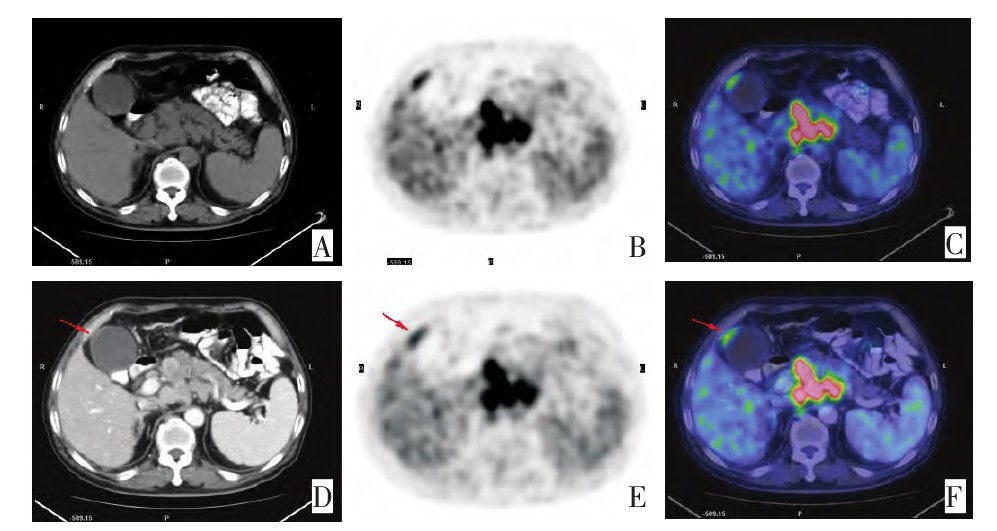
图 1 PET/CT与增强CT联合应用诊断胆囊癌
Figure 1. Combined diagnosis with PET/CT and enhanced CT scan ofgallbladder carcinoma
A man, 64 years old, Figure A(unenhanced CT): the bottom of gallbladder slightly thickened; Figure B, E(PET): the bottom of gallbladder abnormal radioactivity concentration; Figure C(unenhanced PET/CT): suspicious gallbladder; Figure D(enhanced CT): the gallbladder bottom irregular significantly enhanced; Figure F(enhanced PET/CT): gallbladder cancer. Surgical pathology was moderately differentiated adenocarcinoma
表 1 PET/CT与增强CT、B超、MRI诊断胆囊癌的最终结果
例 Table 1. Final diagnostic results of PET/CT, enhanced CT scan, B ultrasound, and MRI in gallbladder carcinoma

表 2 比较PET/CT与其他影像学方法对胆囊癌诊断的价值
例(%) Table 2. Comparison of the diagnostic values between PET/CT and the other imaging methods for gallbladder carcinoma

-
[1] 韩建勋. 胆囊癌的影像学诊断方法与进展[J]. 山西医药杂志, 2012, 41(8): 782-784. doi: 10.3969/j.issn.0253-9926.2012.08.018 [2] 耿金宏, 袁金凤, 吴鸣. 胆囊癌流行分布及高危因素探讨研究[J]. 临床和实验医学杂志, 2012, 11(16): 1336-1337. doi: 10.3969/j.issn.1671-4695.2012.16.043 [3] 李毅红, 张建, 卫建国. 18F-FDG PET/CT对胆囊癌分期、治疗后再分期诊断的价值[J]. 临床影像技术, 2009, 24(09): 74-76. https://cdmd.cnki.com.cn/Article/CDMD-10161-1014331474.htm [4] 梁廷波, 白雪莉. 胆囊癌分期临床意义及评价[J]. 中国实用外科杂志, 2011, 31(3): 194-197. https://www.cnki.com.cn/Article/CJFDTOTAL-ZGWK201103005.htm [5] 石景森, 郑见宝, 孙学军. 提高胆囊癌早期诊断率的临床思维[J]. 肝胆胰外科杂志, 2012, 24(6): 441-443. doi: 10.3969/j.issn.1007-1954.2012.06.001 [6] Makino I, Yamaguchi T, Sato N, et al. Xanthogranulomatous cholecystitis mimicking gallbladder carcinoma with a false-positive result on fluorodeoxyglucose PET[J]. World J Gastroenterol 2009Aug7, 15(29): 3691-3693. [7] Anderson CD, Rice MH, Pinson CW, et al. FluorodeoxyglucosePET Imaging in the Evaluation of Gallbladder Carcinoma andCholangiocarcinoma[J]. J Gastrointest Surg, 2004, 8(1): 90-97. doi: 10.1016/j.gassur.2003.10.003 [8] Koh T, Taniguchi H, Yamaguchi A, et al. Differential Diagnosis ofGallbladder Cancer Using Positron Emission T omography withFluorine-18-Labeled Fluoro-Deoxyglucose(FDG-PET)[J]. J SurgOncol, 2003, 84(2): 74-81. [9] 周信远, 王琛. 原发性胆囊癌的诊疗进展[J]. 中国医学工程, 2012, 20(6): 183-188. https://www.cnki.com.cn/Article/CJFDTOTAL-YCGC201206139.htm [10] Ramos-Font, Santiaqo CA, Rodriquez FA, et al. Gallbladder cancerstaging with 18F-FDG PET/CT[J]. Rev Esp Med Nucl, 2009, 28(2): 74-77. [11] Kim JY, Lee TY, Lee TY, et al. Clinical role of 18F-FDG PET/CTin suspected and potentially operable cholangiocarcinoma: a prospective study compared with conventional imaging[J]. Am J Gastroenterol, 2008, 103(5): 1145-1151. [12] 刘迎春. 探讨螺旋CT增强扫描在胆囊癌诊断中的价值[J]. 中国医药指南, 2011, 9(27): 200-201. doi: 10.3969/j.issn.1671-8194.2011.27.154 [13] 陆健美. CT对原发性胆囊癌的诊断价值[J]. 牡丹江医学院报, 2012, 33(1): 19-21. https://www.cnki.com.cn/Article/CJFDTOTAL-CTMR201409003.htm [14] 杜训松, 魏渝清. 厚壁型胆囊癌与慢性胆囊炎的CT鉴别诊断[J]. 临床放射学杂志, 2008, 27(10): 1331-1334. doi: 10.3969/j.issn.1001-9324.2008.10.013 [15] 张水兴, 梁长虹, 罗海营. 增强MRI及MRCP术前评估肝门胆管癌的可切除性[J]. 磁共振成像, 2010, 1(3): 200-203. https://www.cnki.com.cn/Article/CJFDTOTAL-CGZC201003014.htm -

 下载:
下载:
 点击查看大图
计量
- 文章访问数: 18
- HTML全文浏览量: 22
- PDF下载量: 1
- 被引次数: 0
