
Epithelioid angiomyolipoma of the kidney: a report of two cases and literature review
-
摘要:
目的 探讨肾上皮样血管平滑肌脂肪瘤(epithelial angiomyolipoma, EAML)的临床病理特征, 进一步认识本疾病。 方法 回顾性研究2002年7月至2012年8月2例肾EAML患者的临床及病理资料并进行随访, 结合复习文献, 对其临床病理特点、生物学行为及预后进行分析。 结果 2例患者都为女性, 平均年龄47.5岁, 均为左肾EAML。无明显阳性体征, 2例在B超和CT均发现肾占位, 其中1例CT强化不均匀, 1例增强检查呈渐进性强化, 延迟期强化程度略减低。分别行左肾部分切除术和根治性左肾切除术。组织形态学的主要特征均呈明显的上皮样分化。 结论 分析结果提示: EAML生物学行为与经典型肾AML不同特征在于浸润性生长方式、细胞学异型性, CT对其诊断有重要意义, 但病理结合免疫组化能够确诊, 手术是主要的治疗方法, 其预后大多数良好。 -
关键词:
- 肾脏 /
- 上皮样血管平滑肌脂肪瘤 /
- 诊断 /
- 治疗 /
- 预后
Abstract:Objective This study aimed to analyze the histopathological and immunohistological characteristics, biologic behaviors, and prognosis of two cases of recurrent renal epithelioid angiomyolipoma(EAML). Methods Two cases of EAML were identified between July 2002 and August 2012 in our institute. Review of literature, as well as observation and analysis of clinical pathological features, biological behavior, and prognosis, was conducted. Results The two cases involved in this study were both females whose average age was 47.5. The EAML both occurred on the patients' left kidney. Through computed tomography(CT) one of them was found to show non-uniform enhancement. Enhanced CT showed progressive strengthening and slight reduction in size. Partial nephrectomy of the left kidney and radical left nephrectomy were used for treatment. The epithelioid cells were arranged in nests or in sheets; the epithelioid cells were large with round or polygon shape. Conclusion he primary difference between epithelioid AML and classic AML of the kidney is the manifestation of rare biological behaviors of invasive proliferation and histological features of atypical hyperplasia. CT is important in making a definite diagnosis by pathology and immunohistochemistry. Surgery is the main treatment method, and no standard prognosis exists to date. -
Key words:
- kidney /
- epithelioid angiomyolipoma /
- diagnosis /
- treatment /
- prognosis
-

图 1 病例1CT示:左肾肿物突出于轮廓外,密度欠均匀,增强后明显不均匀强化
Figure 1. CT examination of case1: Left renal neoplasm outstanding in profile outside, density under uniform, the enhanced obviously uneven reinforcement

图 2 病例1免疫组化P53呈阳性表达(免疫组化法×400)
Figure 2. Immunochemical staining of p53 of case 1(IHC×400)

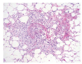
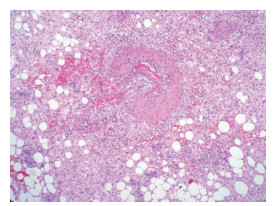
图 3 病例1术后镜下示:肿瘤的主要成分为上皮样细胞,呈巢状或片状排列,肿瘤细胞呈圆形或多角形,核大,核仁清楚(H&E×400)
Figure 3. Postoperative H&E pathology of case 1. The main components of the tumor were epithelioid cells, in round or polygonal shape, arranged in nests or flake, with large and clear nucleolus(H&E×400)
-
[1] John NE, Guido S, Jonathan I, et al. World health organization classif ication of tumours: Pathology and genetics of tumours of the urinary system and male genital organs[M]. Lyon: IARC Press, 2004: 65-69. [2] John WB. Angiolipomyosarcoma of kidney (malignant hamartomatous-angiomyolipoma) in a case with solitary met astasis from bronchogeniccarcinoma[J]. Cancer, 1955, 8(4): 759-763. doi: 10.1002/1097-0142(1955)8:4<759::AID-CNCR2820080418>3.0.CO;2-L [3] Mai KT, Perkins DG, Collins JP. Epithelioid cell variant of renal angiomyolipoma[J]. Histopathology, 1996, 28(3): 277-280. doi: 10.1046/j.1365-2559.1996.d01-421.x [4] Huang KH, Huang CY, Chuang SD, et al. Malignant epothelioid angiomyolipoma of the kidney[J]. J Formos Med Assoc, 2007, 106(2): S51-S54. doi: 10.1016/S0929-6646(09)60353-3 [5] Andrew JG, Tiina S, John RW, et al. Clonality of tuberous sclerosis hamartomas shown by non-random X-chromosome inactivation[J]. Hum Genet, 1996, 97(2): 240-243. doi: 10.1007/BF02265273 [6] Chandrasoma S, Moatamed N, Chang A, et al. Angiomyolipoma of the kidney: expanding disease spectrum demonstrated by 3 cases[J]. Appl Immunohistochem Mole Morphol, 2004, 12(3): 277-283. doi: 10.1097/00129039-200409000-00016 [7] Delgado R, De LB, Albores SJ. Atypical angiomyolipoma of the kidney: a distinct morphologie variant that is easily confused with a variety of malignant neoplasms[J]. Cancer, 1998, 83(8): 1581-1592. doi: 10.1002/(SICI)1097-0142(19981015)83:8<1581::AID-CNCR13>3.0.CO;2-R [8] Eble JN, Amin MB, Young RH. Epithelioid angiomyolipoma of the kidney: a report of five cases with a prominent and diagnostically confusing epithelioid smooth muscle component[J]. Am J Surg Pathol, 1997, 21(10): 1123-1130. doi: 10.1097/00000478-199710000-00001 [9] Kawaguchi K, Oda Y, Nakanishi K, et al. Malignant Transformation of Renal Angiomyolipoma: A Case Report[J]. Am J of Surg Pathol, 2002, 26(4): 523-529. doi: 10.1097/00000478-200204000-00017 [10] Morioka M, Kinugawa K, Funabiki S, et al. Monotypic epithelioid angiomyolipoma of the kidney: a case report[J]. Int Urol Nephrol, 2006, 13(9): 1240-1242. https://pubmed.ncbi.nlm.nih.gov/16984561/#:~:text=Monotypic%20epithelioid%20angiomyolipoma%20of%20the%20kidney%3A%20a%20case,renal%20mass%20without%20evidence%20of%20tuberous%20sclerosis%20complex. [11] Inci O, Kaplan M, Yalcin O, et al. Renal angiomyolipoma with malignant transformation, simultaneous occurrence with malignity and other complex clinical situations[J]. Int Urol Nephrol, 2006, 38(10): 417-426. doi: 10.1007/s11255-005-4756-2 [12] 单世馨. 不典型肾血管平滑肌脂肪瘤12例CT误诊分析[J]. 中国误诊学杂志, 2008, 8(3): 606-607. doi: 10.3969/j.issn.1009-6647.2008.03.085 [13] Khaitan A, Hemal AK, Seth A, et al. Management of renal angiomyolipoma in complex clinical situations[J]. Urol Int, 2001, 67(1): 28-33. doi: 10.1159/000050940 [14] Belanger EC, Dhamanaskar PK, Mai KT. Epithelioid angiomyolipoma of the kidney mimicking renal sarcoma[J]. Histopathology, 2005, 47(4): 433-435. doi: 10.1111/j.1365-2559.2005.02134.x [15] Achim A, Jungbluth, Iversen K, et al. Expression of melanocyte associated m arkers gp100 and Melan-A /MART-1 in angiomyolipomas: An immunohistochemical and RT-PCR analysis[J]. Virchows Arch, 1999, 434(5): 429-435. doi: 10.1007/s004280050362 -

 下载:
下载:

 点击查看大图

计量
- 文章访问数: 42
- HTML全文浏览量: 2
- PDF下载量: 0
- 被引次数: 0
