
-
-

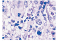
图 1 数量不等的霍奇金细胞并混杂大量的炎性反应性背景细胞(H&E ×400)
Figure 1. Hodgkin's cells, with alot of reactive inflammatory cells(H&E staining ×400)
-
[1] Wang SA, Rahemtullah A, Faquin WC, et al. Hodgkin's lympho ma of the thyroid: a clinicopathologic study of five cases and re view of the literature. Mod Pathol. 2005 Dec; 18 (12): 1577-84. [2] Aiken, A. H. A. H., & amp; Glastonbury, C. C. Imaging hodgkin andnon-hodgkin lymphoma in the head and neck. Radiologic Clinicsof North America, 2008;46 (2), 363-78, ix-x. -

 下载:
下载:
 点击查看大图

图(2)
计量
- 文章访问数: 9
- HTML全文浏览量: 22
- PDF下载量: 0
- 被引次数: 0
