
EphA2/ephrinA1 expression in human malignant gliomas and its relationship with angiogenesis
-
摘要:
目的 检测酪氨酸激酶受体EphA2及其配体EphrinA1在恶性脑胶质瘤中的表达,并探讨其与胶质瘤血管生成的关系。 方法 采用免疫组织化学S-P法检测62例胶质瘤组织及8例正常脑组织中EphA2、EphrinA1的表达情况,并采用CD105抗体标记微血管内皮细胞,计算微血管密度(microvesseldensity,MVD)。探讨EphA2、EphrinA1的表达水平与胶质瘤的微血管生成之间可能存在的关系。 结果 EphA2、MVD在胶质瘤中阳性表达明显高于正常脑组织,二者差异显著(P<0.01)。而且随着肿瘤恶性程度增加,EphA2及MVD蛋白染色强度和阳性细胞数均明显升高,高级别脑胶质瘤(Ⅲ~Ⅳ)中EphA2及MVD强阳性表达明显高于低级别胶质瘤组,差异有显著统计学意义(P<0.01)。EphrinA1则具有相反的趋势,在大多数恶性脑胶质瘤中呈低水平表达,在低级别胶质瘤和正常组织中呈较高水平表达,且胶质瘤级别越低EphrinA1表达越高。EphA2表达与MVD呈显著正相关(r=0.713,P<0.01),EphrinA1表达与MVD呈显著负相关(r=-0.772,P<0.01),EphA2的表达与EphrinA1呈显著负相关(r=-0.912,P<0.01)。 结论 EphA2在恶性脑胶质瘤中特异性高表达、EphrinA1特异性低表达与胶质瘤的侵袭性和恶性程度密切相关,且EphA2过表达和EphrinA1的缺陷表达可能协同促进胶质瘤组织新生血管的生成,在胶质瘤的发病及恶性进展中发挥重要作用。 -
关键词:
- 脑胶质瘤 /
- EphA2 /
- EphrinA1 /
- CD105微血管密度 /
- 血管生成
Abstract:Objective To investigate the expressions and significance of tyrosine kinase receptor EphA2 and its ligand ephrinA1 in human malignant gliomas and their correlation with tumor angiogenesis. Methods The expressions of EphA2, ephrinA1, and CD105-stained microvessel density (MVD) were detected via immunohistochemical assay in 62 glioma tissues and 8 normal brain tissues. The correlation between EphA2 and ephrinA1 expression and microvessel counts in the glioma tissues were assessed. Results Immunohistochemical staining results revealed that variable levels of EphA2 and MVD expression were significantly higher than that of the normal brain samples. Statistical difference was observed in EphA2 and MVD expressions between human gliomas and normal brain samples (P<0.01). The positive rate of EphA2 and MVD expressions was significantly higher in high-grade gliomas (WHO Ⅲ-Ⅳ) than that in low-grade gliomas (WHO Ⅰ-Ⅱ)(P<0.01). EphrinA1 was expressed at low levels in most malignant gliomas, and the increased ephrinA1 expression was associated with lower-grade histology. MVD was significantly positively correlated with EphA2 expression (r=0.713, P<0.01) and significantly negatively correlated with ephrinA1 expression (r=-0. 772, P<0.01). EphA2 was significantly negatively correlated with ephrinA1 expression (r=-0.912, P<0.01). Conclusion Specifically over-expressed EphA2 and its low-expressed ligand ephrinA1 in malignant gliomas may be closely correlated with the invasion and malignant degree of gliomas. Cooperation is involved in the angiogenesis and has an important function in the initiation and progression of gliomas. -
Key words:
- gliomas /
- EphA2 /
- EphrinA1 /
- CD105-MVD /
- angiogenesis
-

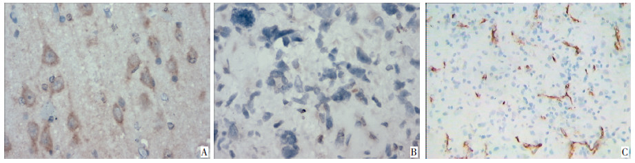
图 1 EphA2、EphrinA1、CD105在高级别胶质瘤组织中的表达(S-P ×400)
Figure 1. Expression of EphA2, EphrinA1, and CD105 in high-grade gliomas(S-P ×400)
A: EphA2;B: EphrinA1;C: CD105

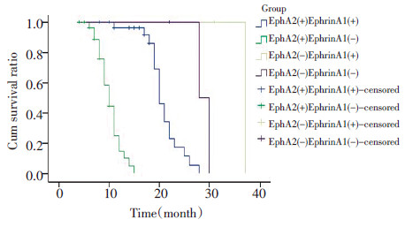
图 2 不同EphA2与EphrinA1表达脑胶质瘤患者的Kaplan-Meier生存曲线
Figure 2. Kaplan-Meier survival curves of patients with positive and neg⁃ ative EphA2 and EphrinA1 expression
表 1 EphA2、EphrinA1和CD105-MVD在正常脑组织及胶质瘤组织中的表达
Table 1. Expression of EphA2, EphrinA1, and CD105-MVD in gliomas and normal brain based on immunohistochemistry results

表 2 EphA2、EphrinA1分别和MVD在胶质瘤中表达的相关性
Table 2. Correlation among the expression of EphA2, EphrinA1, and MVD in gliomas

表 3 EphA2和EphrinA1在胶质瘤中表达的相关性
Table 3. Correlation between the expression of EphA2 and EphrinA1 in gliomas

-
[1] 郑俊青, 安聪娟, 方艳伟. EphA2/ephrinA1与人脑胶质瘤发生的关系及靶向治疗潜能探讨[J]. 中国肿瘤临床, 2013, 40(8): 486-488. doi: 10.3969/j.issn.1000-8179.2013.08.014 [2] Tanaka F, Otake Y, Yanagihara K, et al. Evaluation of angioagenesis in non-Small cell lung cancer: comparison between anti-CD34 antibody and anti-CD105 antibody[J]. Clin Cancer Res, 2001, 7(11): 3410-3415. [3] Merritt WM, Kamat AA, Hwang JY, et al. Clinical and biological impact of EphA2 overexpression and angiogenesis in endometrial cancer[J]. Cancer Biol Ther, 2011, 10(12): 1306-1314. [4] Weidner N. Current pathologic methods for measuring intratumoral microvessel density with in breast carcinoma and other solid tumors[J]. Breast Cancer Res Treat, 1995, 36(2): 169-180. doi: 10.1007/BF00666038 [5] 刘辉, 郭建文, 刘江惠, 等. EphA2/EphrinA1在胃癌中的表达及其意义[J]. 中国肿瘤临床, 2008, 35(10): 573-577. [6] 徐金升, 王悦芬, 李亚林, 等. 肾癌中EphA2/EphrinA1和E-cadherin的表达及其意义[J]. 中国肿瘤临床, 2008, 35(22): 1276-1280. [7] Hochgräfe F, Zhang L, O'Toole SA, et al. Tyrosine phosphorylation profiling reveals the signaling network characteristics of Basal breast cancer cells[J]. Cancer Res, 2010, 70(22): 9391-9401. doi: 10.1158/0008-5472.CAN-10-0911 [8] Zhou N, Zhao WD, Liu DX, et al. Inactivation of EphA2 promotes tight junction formation and impairs angiogenesis in brain endothelial cells[J]. Microvasc Res, 2011, 82(2): 113-121. doi: 10.1016/j.mvr.2011.06.005 [9] Wykosky J, Palma E, Gibo DM, et al. Soluble monomeric ephrinA1 is released from tumor cells and is a functional ligand for the EphA2 receptor[J]. Oncogene, 2008, 27(58): 7260-7273. doi: 10.1038/onc.2008.328 [10] Korkolopoulou P, Patsouris E, Kavantzas N, et al. Prognostic implications of microvessel morphometry in diffuse astrocytic neoplasms [J]. Neuropathol Appl Neurobiol, 2002, 28(1): 57-66. doi: 10.1046/j.1365-2990.2002.00367.x [11] Folkman J. Angiogenesis in cancer, vascular, rheumat oid and other diseases[J]. Nature Med, 1995, 1(1): 27-31. doi: 10.1038/nm0195-27 [12] Hess AR, Seftor EA, Gardner LM, et al. Molecular regulation of tumor cell vasculogenic mimicry by tyrosinephosphorylation: role of epithelial cell kinase(Eck/EphA2)[J]. Cancer Res, 2001, 61(8): 3250-3255. [13] Ogawa K, Pasqualini R, Lindberg RA, et al. The ephrin-A1 ligand and its receptor, EphA2, are expressed during tumor neovascularization[J]. Oncogene, 2000, 19(52): 6043-6052. doi: 10.1038/sj.onc.1204004 [14] Larsen AB, Stockhausen MT, Poulsen HS. Cell adhesion and EGFR activation regulate EphA2 expression in cancer[J]. Cell Signal, 2010, 22(4): 636-644. doi: 10.1016/j.cellsig.2009.11.018 [15] Xu JS, Zhang JX, Geng TH, et al. Expression of EphA2 and ephrinA1 in human renal cell carcinoma and its relationship with angiogenesis[J]. Zhonghua Zhong Liu Za Zhi, 2009, 31(6): 438-441. -

 下载:
下载:
 点击查看大图
计量
- 文章访问数: 50
- HTML全文浏览量: 14
- PDF下载量: 0
- 被引次数: 0
