
-
摘要:
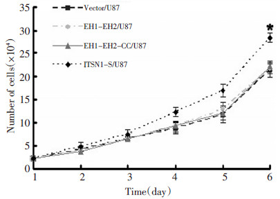
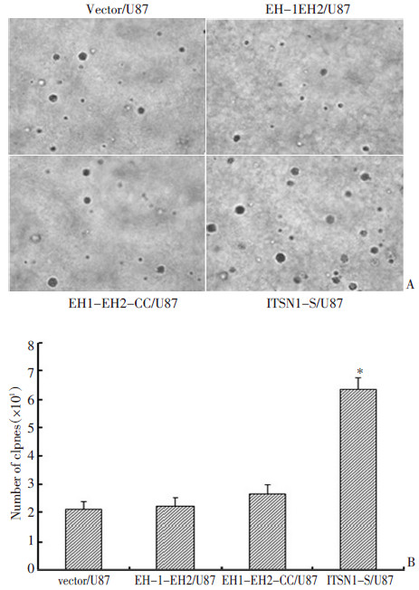
目的 观察ITSN1-S的SH3功能域对恶性胶质瘤细胞U87增殖能力的影响,并探讨其相关分子机制。 方法 构建标记mGFP荧光的慢病毒表达质粒PCDH-CMV-MCS-EF1-Puro。将ITSN1-S的不同片段的PCR产物:EH1-EH2、EH1-EH2-CC、ITSN1-S,分别连接到慢病毒表达质粒载体中。用HEK-293T细胞分别包装上述四种质粒的慢病毒后感染U87细胞,嘌呤霉素(puro)筛选出稳定表达的细胞,分别命名为vector/U87、EH1-EH2/U87、EH1-EH2-CC/U87、ITSN1-S/U87。Western blot检测目的蛋白的表达,增殖实验和软琼脂克隆形成实验检测各组胶质瘤细胞的增殖能力。 结果 增殖实验和软琼脂克隆形成实验显示与vector/U87、EH1-EH2/U87、EH1-EH2-CC/U87细胞相比较,ITSN1-S/U87细胞的增殖能力显著增强(P < 0.05),但vector/U87、EH1-EH2/U87、EH1-EH2-CC/U87三组细胞之间的增殖能力差异无统计学意义(P>0.05)。增殖实验结果显示第6天时,ITSN1-S/ U87、EH1-EH2-CC/U87、EH1-EH2/U87、vector/U87细胞数分别为(29.16±1.19)×104、(22.82±0.94)×104、(22.17±0.90)×104、(21.93± 1.15)×104个;软琼脂克隆形成实验表明第21d时,ITSN1-S/U87克隆形成数高达(6.37±0.41)×103个,而EH1-EH2-CC/U87、EH1-EH2/U87和vector/U87克隆形成数分别为(2.65±0.34)×103、(2.23±0.31)×103和(2.1±0.29)×103个。 结论 过表达ITSN1-S能显著提高胶质瘤细胞U87的增殖能力,这种促进细胞增殖的作用可能是通过SH3功能域来实现的。 Abstract:Objective To investigate the functions of the ITSN1-S SH3 domains in U87 glioblastoma cell proliferation and to determine the underlying molecular mechanism. Methods A recombinant lentiviral vector with an mGFP label was constructed. EH1-EH2, EH1-EH2-CC, and ITSN1-S genes were amplified using polymerase chain reaction and then cloned into recombinant lentiviral vectors. The four lentiviral plasmids were packaged using HEK 293T cells and subsequently used to infect U87 cells. Stable cells were screened using puromycin and separately labeled as vector/U87, EH1-EH2/U87, EH1-EH2-CC/U87, and ITSN1-S/U87. Western blotting was used to detect the expression of each protein. Proliferation and soft agar assays were performed to detect cell proliferation. Results In the proliferation and soft agar assays, the proliferation capacity of the ITSN1-S/U87 cells was clearly enhanced compared with those of the vector/U87, EH1-EH2/U87, and EH1-EH2-CC/U87 cells (P < 0.05). Moreover, the proliferation capacity of the latter three groups showed no observable difference (P>0.05). On the 6th day, the vector/U87, EH1-EH2/U87, EH1-EH2-CC/U87, and ITSN1-S/U87 cell numbers were (29.16±1.19)×104, (22.82±0.94)×104, (22.17±0.90)×104, and (21.93±1.15)×104, respectively. On the 21st day, the number of colony formation in vector/U87, EH1-EH2/U87, EH1-EH2-CC/U87, and ITSN1-S/U87 was (6.37±0.41)×103, (2.65±0.34)×103, (2.23±0.31)×103, and (2.1±0.29)×103, respectively. Conclusion ITSN1-S overexpression significantly promotes U87 cell proliferation. Specifically, the SH3 domains possibly serve vital functions in glioma cell proliferation. -
Key words:
- glioma cell U87 /
- ITSN1-S /
- proliferation
-

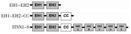
图 1 EH1-EH2、EH1-EH2-CC和ITSN1-S全长三种过表达质粒结构模式图
Figure 1. Structural model of the overexpressed plasmids EH1-EH2, EH1-EH2-CC, and ITSN1-S

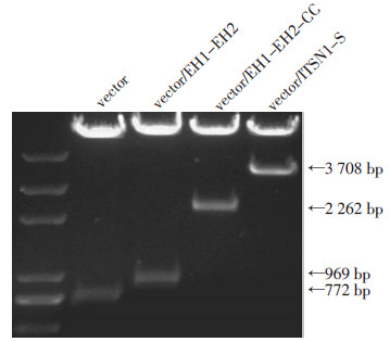
图 2 四种重组质粒的酶切鉴定结果
Figure 2. Four recombinant plasmids are confirmed by enzyme digestion

图 3 Western blot检测EH1-EH2、EH1-EH2-CC和ITSN1-S在U87细胞中的表达情况
Figure 3. EH1-EH2, EH1-EH2-CC, and ITSN1-S expression in U87 cells as determined by Western blot

图 4 EH1-EH2、EH1-EH2-CC和ITSN1-S的表达水平升高后,细胞增殖实验检测四组细胞增殖能力的差异(*p < 0.05)
Figure 4. Proliferation assay results showing the differences in EH1-EH2, EH1-EH2-CC, and ITSN1-S cell proliferation activity after promotion of EH1-EH2, EH1-EH2-CC, and ITSN1-S expression, respectively

图 5 EH1-EH2、EH1-EH2-CC和ITSN1-S的表达水平升高后,软琼脂克隆形成实验检测四组细胞增殖能力的差异(*P < 0.05)
Figure 5. Effect of EH1-EH2, EH1-EH2-CC ITSN1-S expression on U87 proliferation
A: Soft agar assay results showing the differences in EH1-EH2, EH1-EH2-CC, and ITSN1-S cell proliferation activity after promotion of EH1-EH2, EH1-EH2-CC and ITSN1-S expression, respectively; B: Sta⁃ tistic chart of the soft agar assay
-
[1] Miller CR, Perry A. Glioblastoma[J]. Arch Pathol Lab Med, 2007, 131(3): 397-406. doi: 10.5858/2007-131-397-G [2] Predescu SA, Predescu DN, Knezevic I, et al. Intersectin-1s regulates the mitochondrial apoptotic pathway in endothelial cells[J]. J Biol Chem, 2007, 282(23): 17166-17178. doi: 10.1074/jbc.M608996200 [3] Ma Y, Wang B, Li W, et al. Reduction of intersectin1-s induced apoptosis of human glioblastoma cells[J]. Brain Res, 2010, 1351: 222-228. doi: 10.1016/j.brainres.2010.05.028 [4] Ma YJ, Wang B, Li W, et al. Intersectin1-s is involved in migration and invasion of human gliomacells[J]. J Neurosc Res, 2011, 89 (7): 1079-1090. doi: 10.1002/jnr.22616 [5] 王冰冰, 牛瑞芳, 马勇杰. ITSN1-S对胶质瘤细胞凋亡的作用研究[J]. 中国肿瘤临床, 2010, 37(11): 608-610. doi: 10.3969/j.issn.1000-8179.2010.11.003 [6] Pucharcos C, Estivill X, de la Luna S. Intersectin 2, a new multimodular protein involved in clathrin-mediated endocytosis[J]. FEBS Lett, 2000, 478(1-2): 43-51. doi: 10.1016/S0014-5793(00)01793-2 [7] Okamoto M, Schoch S, Sudhof TC. EHSH1/intersectin, a protein that contains EH and SH3 domains and binds to dynamin and SNAP-25. A protein connection between exocytosis and endocytosis[J]? J Biol Chem, 1999, 274(26): 18446-18454. doi: 10.1074/jbc.274.26.18446 [8] Hussain NK, Jenna S, Glogauer M, et al. Endocytic protein intersectin-l regulates actin assembly via Cdc42 and N-WASP[J]. Nat Cell Biol, 2001, 3(10): 927-32. doi: 10.1038/ncb1001-927 [9] Das M, Scappini E, Martin NP, et al. Regulation of neuron survival through an intersectin-phosphoinositide 3'-kinase C2beta-AKT pathway[J]. Mol Cell Biol, 2007, 27(22): 7906-7917. doi: 10.1128/MCB.01369-07 [10] Predescu SA, Predescu DN, Knezevic I, et al. Intersectin-1s regulates the mitochondrial apoptotic pathway in endothelial cells[J]. J Biol Chem, 2007, 282(23): 17166-1778. doi: 10.1074/jbc.M608996200 [11] Tsyba L, Nikolaienko O, Dergai O, et al. Intersectin multidomain adaptor proteins: regulation of functional diversity[J]. Gene, 2011, 473(2): 67-75. doi: 10.1016/j.gene.2010.11.016 [12] Ma YJ, Okamoto M, Gu F, et al. Neuronal distribution of EHSH1/intersectin: molecular linker between clathrin-mediated endocytosis and signaling pathways[J]. J Neurosci Res, 2003, 71(4): 468-477. doi: 10.1002/jnr.10500 [13] Wheeler M, Domin J. The N-terminus of phosphoinositide 3-kinase-C2beta regulates lipid kinase activity and binding to clathrin [J]. J Cell Physiol, 2006, 206(3): 586-593. doi: 10.1002/jcp.20507 [14] Mohney RP, Das M, Bivona TG, et al. Intersectin activates Ras but stimulates transcription through an independent pathway involving JNK[J]. J Biol Chem, 2003, 278(47): 47038- 47045. doi: 10.1074/jbc.M303895200 [15] 李智慧, 谷峰, 马勇杰. Intersectin蛋白在细胞信号通路中的作用研究进展[J]. 中华实验外科杂志, 2012, 29(1): 148-149. doi: 10.3760/cma.j.issn.1001-9030.2012.01.061 -

 下载:
下载:

 点击查看大图
计量
- 文章访问数: 24
- HTML全文浏览量: 5
- PDF下载量: 0
- 被引次数: 0
