
Expression and clinical significances of caveolin-1 in pleomorphic adenoma and paraneoplastic tissues of parotid gland
-
摘要:
目的 探讨窖蛋白-1(Caveolin-1,Cav-1)在腮腺多形性腺瘤(pleomorphic adenoma,PA)中的表达及临床意义。 方法 采用免疫组织化学法检测了9例正常腮腺、33例原发性PA距离瘤体中心(0 cm)及瘤旁组织中(分别距瘤体中心0.5、1.0、1.5和2.0 cm)Cav-1的表达,并进行统计学分析。 结果 Cav-1在所有正常腮腺样本(100%)均有表达,其主要在导管系统细胞质和细胞膜中表达;除6例距瘤体中心0.5 cm和1例距瘤体中心1.0 cm的样本外,其余瘤旁样本均有Cav-1表达,其表达模式基本上与正常腮腺类似;在PA中,有10例样本(30.30%)表达Cav-1,且在细胞核中显著表达。Cav-1在多形性腺瘤肿瘤组织中的表达最低,并随着与瘤体中心距离的远近,其表达量逐渐升高。Cav-1的表达在瘤体与瘤旁组织间均存在明显差异(P < 0.05),但瘤旁组织1.0、1.5和2.0 cm间Cav-1表达无显著性差异(P>0.05)。 结论 在腮腺多形性腺瘤发生过程中,Cav-1中表达下调并出现了从细胞质、细胞膜到细胞核的转位现象。上述结果表明Cav-1在多形性腺瘤的发生发展中可能起着重要作用,其表达水平的变化可作为判定手术安全边缘的科学依据之一。 -
关键词:
- Caveolin-1 /
- 多形性腺瘤 /
- 腮腺 /
- 手术切缘
Abstract:Objective This study was conducted to explore the expression and clinical significance of caveolin-1 (Cav-1) in pleomorphic adenoma and paraneoplastic tissues of the parotid gland. Methods Immunohistochemistry was used to detect Cav-1 expression in normal parotid glands, pleomorphic adenoma (0 cm), and paraneoplastic tissues (0.5, 1.0, 1.5, and 2.0 cm from the pleomorphic adenoma). Results Cav-1 was expressed in all normal parotid glands and was mainly expressed in the cytoplasm and plasma membrane of duct systems. Except for six samples 0.5 cm away and one sample 1.0 cm away from the pleomorphic adenoma, Cav-1 was expressed in other adjacent normal tissues, and the expression pattern of Cav-1 was similar to that in normal tissues. In pleomorphic adenoma, Cav-1 only existed in seven samples (accounting for 30.30%) and was prominently expressed in the nucleus. The expression level of Cav-1 was lowest in pleomorphic adenoma and progressively increased with increasing distance from the tumor. A difference in Cav-1 expression was detected between pleomorphic adenoma (0 cm) and paraneoplastic tissues (0.5, 1.0, 1.5, and 2.0 cm from the tumor, P < 0.05). However, no statistical significance existed between paraneoplastic tissues at 1.0, 1.5, and 2.0 cm from the tumor (P> 0.05). Conclusion During the development of pleomorphic adenoma, the expression level of Cav-1 progressively decreased, and translocation of Cav-1 expression from cytoplasm and plasma membrane to nucleus occurred. Cav-1 may have an important role in the progress of pleomorphic adenoma, and the variation in the expression level of Cav-1 may be used as a scientific evidence to determine the safe surgical margin of pleomorphic adenoma. -
Key words:
- Caveolin-1 /
- pleomorphic adenoma /
- parotid gland /
- surgical margins
-

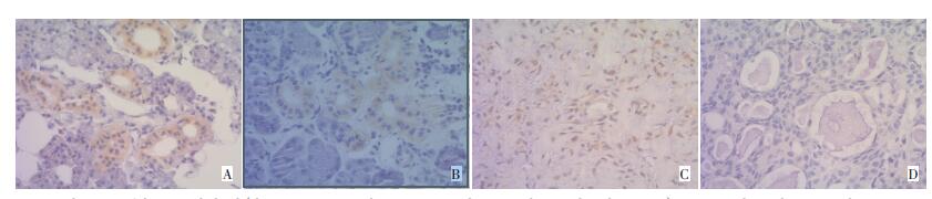
图 1 Cav-1在正常腮腺、多形性腺瘤及瘤旁组织中的表达(H&E×200)
A. Normal tissues of the parotid gland(the arrow points to the positive signals in cytoplasm and nuclear areas);B. tissues located a certain distance away from the pleomorphic adenoma(one sample 0.5 cm away from pleomorphic adenoma,the arrow point to the positive signals in cytoplasm and nuclear);C. pleomorphic adenoma with Cav-1 positive signals(the arrow points to the positive signals in the nuclear area);D. pleomorphic adenoma with negative Cav-1 signal
Figure 1. Expression of Cav-1 in normal tissues and tissues located at a certain distance away from pleomorphic adenoma and pleomorphic adenoma of the parotid gland (H&E×200)
表 1 Caveolin-1在正常腮腺、腮腺多形性腺瘤(PA)及瘤旁组织中的表达分布
例 Table 1. Expression of Cav-1 in normal tissues and tissues located at a certain distance away from pleomorphic adenoma and pleomorphic adenoma of the parotid gland
n 
表 2 Caveolin-1在正常腮腺、腮腺多形性腺瘤(PA)及瘤旁组织细胞核中的表达分布
例 Table 2. Nuclear distributions of Cav-1 in normal tissues and tissues located at a certain distance away from pleomorphic adenoma and pleomorphic adenoma of the parotid gland
n 
-
[1] Senetta R, Stella G, Pozzi E, et al. Caveolin-1 as a promoter of tumour spreading: when, how, where and why[J]. J Cell Mol Med, 2013, 17(3):325-336. doi: 10.1111/jcmm.12030 [2] Mo SJ, Yang SL, Cui Z. New glimpses of caveolin-1 functions in embryonic development and human diseases[J]. Front Biol, 2011, 6 (5):367-376. doi: 10.1007/s11515-011-1132-8 [3] Goetz JG, Lajoie P, Wiseman SM, et al. Caveolin-1 in tumor progression: the good, the bad and the ugly[J]. Cancer Metastasis Rev, 2008, 27(4):715-735. doi: 10.1007/s10555-008-9160-9 [4] Schlegel A, Arvan P, Lisanti MP. Caveolin-1 binding to endoplasmic reticulum membranes and entry into the regulated secretory pathway are regulated by serine phosphorylation. Protein sorting at the level of the endoplasmic reticulum[J]. J Biol Chem, 2001, 276(6):4398-4408. doi: 10.1074/jbc.M005448200 [5] Sunaga N, Miyajima K, Suzuki M, et al. Different roles for caveolin-1 in the development of non-small cell lung cancer versus small cell lung cancer[J]. Cancer Res, 2004, 64(12):4277-4285. doi: 10.1158/0008-5472.CAN-03-3941 [6] 李超, 徐义全, 樊晋川.腮腺多形性腺瘤手术切缘研究进展[J].中国肿瘤临床, 2011, 38(4):238-240.Li C, Xu YQ, Fan JC. Advances in the Research of Surgical Margins for Pleomorphic Adenoma of Parotid Gland[J]. Chin J Clin Oncol, 2011, 38(4):238-240. [7] Shi L, Chen XM, Wang L, et al. Expression of caveolin-1 in mucoepidermoid carcinoma of the salivary glands: correlation with vascular endothelial growth factor, microvessel density, and clinical outcome[J]. Cancer, 2007, 109(8):1523-1531. doi: 10.1002/cncr.22573 [8] 史璐, 陈新明, 陈智, 等.涎腺粘液表皮样癌中Caveolin-1、PCNA的表达及意义[J].口腔医学研究, 2011, 27(7):596-599. doi: 10.7666/d.d129092Shi L, Chen XM, Chen Z, et al. Expression and Significant of Caveolin-1 and PCNA in Mucoepidermold Carcinoma of Salivary Glands[J]. Journal of Oral Science Research, 2011, 27(7):596-599. doi: 10.7666/d.d129092 [9] Xia X, Ma Q, Li X, et al. Cytoplasmic p21 is a potential predictor for cisplatin sensitivity in ovarian cancer[J]. BMC Cancer, 2011, 11:399. doi: 10.1186/1471-2407-11-399 [10] Saifo MS, Rempinski DJ, Rustum YM, et al. Targeting the oncogenic protein beta-catenin to enhance chemotherapy outcome against solid human cancers[J]. Mol Cancer, 2010, 9:310. doi: 10.1186/1476-4598-9-310 [11] Lu Z, Ghosh S, Wang Z, et al. Downregulation of caveolin-1 function by EGF leads to the loss of E-cadherin, increased transcriptional activity of beta-catenin, and enhanced tumor cell invasion[J]. Cancer Cell, 2003, 4(6):499-515. doi: 10.1016/S1535-6108(03)00304-0 [12] Masuelli L, Budillon A, Marzocchella L, et al. Caveolin-1 overexpression is associated with simultaneous abnormal expression of the E-cadherin/α-β catenins complex and multiple erbb receptors and with lymph nodes metastasis in head and neck squamous cell carcinomas[J]. J Cell Physiol, 2012, 227(9):3344-3353. doi: 10.1002/jcp.24034 [13] 汤国雄, 朱声荣, 陈卫民, 等.腮腺多形性腺瘤及其恶变中β-连环蛋白、细胞周期蛋白D 1的表达[J].临床口腔医学杂志, 2009, 25(3):131-133. doi: 10.3969/j.issn.1003-1634.2009.03.001Tang GX, Zhu SR, Chen WM, et al. Expression and significance of β-catenin, CyclinDl in pleomorphic adenoma and carcinoma in pleomorphic adenoma of parotid gland[J]. J Clin Stomatol, 2009, 25 (3):131-133. doi: 10.3969/j.issn.1003-1634.2009.03.001 [14] Hu J, Shao S, Song Y, et al. Hepatocyte growth factor induces invasion and migration of ovarian cancer cells by decreasing the expression of E-cadherin, beta-catenin, and caveolin-1[J]. Anat Rec (Hoboken), 2010, 293(7):1134-1139. doi: 10.1002/ar.21147 [15] 莫赛军, 张淑伟, 杨胜利.caveolin-1基因与肿瘤发生相关性的研究进展[J].癌变.畸变.突变, 2012, 24(6):477-479. doi: 10.3969/j.issn.1004-616x.2012.06.019Mo SJ, Zhang SW, Yang SL. Research Advances in the Correlation between caveolin-1 gene and Oncogenesis[J]. Carcinogenesis, Teratogenesis & Mutagenesis, 2012, 24(6):477-479. doi: 10.3969/j.issn.1004-616x.2012.06.019 -

 下载:
下载:
 点击查看大图
计量
- 文章访问数: 7
- HTML全文浏览量: 21
- PDF下载量: 0
- 被引次数: 0
