
Diagnostic value of separated cystic lesion ultrasound and contrast-enhanced ultrasound for multi-locular cystic renal cell carcinoma and cysts
-
摘要:
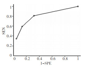
目的 探讨肾脏多房状囊性占位灶内分隔的彩色多普勒以及超声造影表现对多房囊性肾癌及多房肾囊肿的诊断价值。 方法 对53例(共54个病灶)经手术病理证实为多房状囊性肾癌以及多房状肾囊肿的患者进行超声检查,同时对该组24例患者进行超声造影(共24个病灶)检查,采用ROC曲线对病灶内分隔数目、厚度、分隔上的血流以及分隔的超声造影特征进行分析。 结果 通过对本组53例患者共54个病灶内分隔数目、厚度以及分隔上血流的ROC曲线分析,病灶内分隔数目≥5条、3 mm < 厚度≤4 mm及分隔上出现条状血流时其诊断囊性肾癌的特异度较高(分别为86%、95%、86%)。三者曲线下面积显示均具有较高的诊断价值(Az分别为0.7621、0.8331、0.7962)。而分隔数目等于4条,2 mm < 厚度≤3 mm以及分隔出现点状血流时虽可作为诊断的最佳临界值,14例多房状囊性肾癌与10例多房状肾脏囊肿内分隔超声造影开始增强时间分别为(11.2±3.4)s及(18.4±4.5)s,达峰时间分别为(21.7±3.8)s及(37.8±8.0)s,开始消退时间分别为(32.1±4.0)s及(51.3±9.0)s,二者之间比较差异均具有统计学意义(t或t'值分别为4.47、5.90、6.31,P < 0.05)。 结论 多房状肾囊性占位灶内分隔数目、厚度以及分隔上血流超声表现对多房状囊性肾癌诊断具有较高的特异度,ROC曲线显示具有较高诊断价值,病灶内分隔的超声造影表现有助于多房状囊性肾癌与多房状肾囊肿的鉴别。 Abstract:Objective To investigate the diagnostic values of separated renal multi-locular cystic lesions color Doppler ultrasound and contrast-enhanced ultrasound performance in multi-locular cystic renal cell carcinoma and cysts. Methods A total of 53 patients (54 lesions) with multi-locular cystic renal cell carcinoma and cysts were included in the study. The presence of carcinoma and cysts was confirmed via histopathology and tested using ultrasound. Contrast-enhanced ultrasound was applied in 24 (24 lesions) of the total number of patients, and the receiver operating characteristic (ROC) curve was used to analyze the numbers of separation, thickness, and type of blood flow patterns of the lesions. The contrast-enhanced ultrasound characteristics were also analyzed. We analyzed the diagnostic value of the color Doppler ultrasound in the separated renal multilocular cystic lesions and the contrast-enhanced ultrasound performance in multi-locular cystic renal cell carcinoma and cysts. Results Based on the analysis of the ROC curves of the separation number, thickness, and type of the blood flow of the lesions in 53 patients (54 lesions), the diagnostic specificity was relatively higher in the lesions where the separation number was ≥5 strips (86%), the thicknesses were >3 and ≤4 mm (95%), and blood flow was band-like (86%). The areas under the curve of the three indexes were 0.7621, 0.8331, and 0.7962, respectively, which indicate high diagnostic values. The separation number of 4 strips, the thicknesses of >2 and ≤3 mm, and the point-like blood flow could be used as critical values for the diagnosis. The contrast enhancement, enhancement peak, and disappearance were (11.2±3.4), (21.7±3.8), and (32.1±4.0) s in 14 patients with multi-locular cystic renal cell carcinoma and (18.4±4.5), (37.8±8.0), and (51.3±9.0) s in 10 patients with multi-locular renal cysts, with statistically significant differences (t=4.47, t'=5.90, t'=6.31, respectively; P < 0.05). Conclusion The separation number, thickness, and type of blood flow of lesions have relatively higher specificity in multi-locular renal cysts than in multi-locular cystic renal cell carcinoma. The ROC curves show a high diagnostic value. Contrast-enhanced ultrasound of the lesions helped in the differential diagnosis of multi-locular cystic renal cell carcinoma and renal cysts. -

图 1 多房状肾囊肿与多房状囊性肾癌分隔数目比较
Figure 1. Comparison of numbers of separation between multilocular cystic renal cell carcinoma and renal cysts separated

图 2 多房状肾囊肿与多房状囊性肾癌分隔厚度比较
Figure 2. Comparison of thickness of separa⁃ tion between multilocular cystic renal cell carci⁃ noma and renal cysts separated

图 3 多房状肾囊肿与多房状囊性肾癌分隔上血流比较
Figure 3. Comparison of blood flow in the separation between multilocular cystic renal cell carcinoma and renal cysts separated

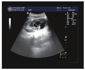
图 4 囊性肾癌患者,囊内更多分隔
Figure 4. Patients with multiple cystic renal cell carcinoma, more separations in cyst

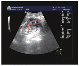
图 5 囊性肾癌患者,囊内分隔较厚并分隔上血流信号丰富
Figure 5. Patients with multiple cystic renal cell carcinoma, more thick separations with abundant blood flow signal

图 6 囊性肾癌患者,超声造影显示囊内分隔快速增强
Figure 6. Patients with multiple cystic renal cell carcinoma, sac separated rapidly enhanced
表 1 多房状囊性肾癌与多房状肾囊肿分隔数目、厚度、血流分布比较表
Table 1. Comparison of number, thickness, and blood flow distribution between multi-locular cystic renal cell carcinoma and cysts

-
[1] Israel GM, Hindman N, Bosniak MA. Evaluation of cystic renal masses: comparison of CT and MR imaging by using the Bosniak classification system[J]. Radiology, 2004, 231(2):365 -371. doi: 10.1148/radiol.2312031025 [2] 王金锐, 苗立英, 崔立刚, 等.超声造影在囊性肾病变中的应用价值[J].中国医学科学院学报, 2008, 30(1)22-26. doi: 10.3321/j.issn:1000-503X.2008.01.005Wang JR, Miao LY, Cui LG, et al. Application of Contrast-en hanced Ultrasound in the Diagnosis of Renal Cystic Lesions[J]. Ac ta Academic Medicinae Sinicae, 2008, 30(1):22-26. doi: 10.3321/j.issn:1000-503X.2008.01.005 [3] 蒋珺, 陈亚青, 朱云开, 等.超声造影结合Bos niak分级诊断囊性肾癌的可行性[J].中国医学影像技术, 2010, 26(3) 549-552.Jiang J, Chen YQ, Zhu YK, et al. Feasibility of Contrast-enhanced Ultrasonography combined with Bosniak Classification in diagnosis of cystic renal cystic carcinoma[J]. China J Med Imaging Technol, 2010, 26(3)549-552. [4] 许小云, 杜联芳, 邢晋放, 等.超声造影在囊性肾癌鉴别诊断中的价值[J].临床超声医学杂志, 2007, 9(11):664-666. doi: 10.3969/j.issn.1008-6978.2007.11.008Xu XY, Du LY, Xin JF, et al. Contrast-enhanced ultrasonography in differential diagnosis of cystic renal cell carcinoma[J]. Journal of Ultrasound in Clin Med, 2007, 9(11):664-666. doi: 10.3969/j.issn.1008-6978.2007.11.008 [5] 黄备建, 王文平, 丁红, 等.超声造影在囊性肾癌诊断中的应用价值[J].中华医学超声杂志.2008;5(4):639-644. doi: 10.3969/j.issn.1672-6448.2008.04.014Huang BJ, Wang WP, Ding H, et al. The value of contrast-en hanced ultrasound in the diagnosis of cystic renal cell carcinoma[J]. Chin J Med Ultrasound, 2008; 5(4):639-644. doi: 10.3969/j.issn.1672-6448.2008.04.014 [6] 蒋珺, 陈亚青, 周永昌.囊性肾癌的超声造影与增强CT对照研究[J].中国医学影像技术, 2008, 24(10):1628-1631. doi: 10.3321/j.issn:1003-3289.2008.10.040 [7] Park BK, Kim B, Kim SH, et al. Assessment of cystic renal masses based on Bosniak classification: comparison of CT and contrast-en hanced US[J]. Eur J Radiol, 2007, 61(2):310-314. doi: 10.1016/j.ejrad.2006.10.004 [8] 陈宇.薛铁, 郝玉芝, 等.超声和超声造影对肾囊性肿物的诊断价值[J].中国肿瘤, 2012, 2l(3):234-236. doi: 10.11735/j.issn.1004-0242.2012.3.A015Chen Y, Xu T, Hao YZ, et al. The diagnostic value of ultrasonogra phy and contrast enhanced ultrasonography for renal cystic mass. china cancer[J]. Chinese Journal of Oncology, 2012, 2l(3):234-236. doi: 10.11735/j.issn.1004-0242.2012.3.A015 [9] Chami L, Lassau N, Malka D, et al. Benefits of contrast-enhanced ultrasonography for the detection of liver lesions: comparison with histologic findings[J]. AJR Am J Roentgenol, 2008, 190(3):683-90. doi: 10.2214/AJR.07.2295 [10] Hatanaka K, Kudo M, Minami Y, et al. Differential diagnosis of he patic tumors: Va lue of contrast-enhanced harmonic ultrasonogra phy using the newly developed contrast agent[J]. Sonazoid Intervi rology, 2008, 51(1):61-69. doi: 10.1159/000122600 [11] Klein D, Jenett M, Gassel H, et al. Quantitative dynamic con trast-enhanced sonography of hepatic tumors[J]. European Radiolo gy, 2004, 14(6):1082-1091. doi: 10.1007/s00330-004-2299-z [12] Romanini L, Passamonti M, Aiani L, et al. Economic assessment of contrast-enhanced ultrasonography for evaluation of focal liver le sions: A multicentre Italian experience[J]. Eur Radiol, 2007, 17(6): 99-106. http://cn.bing.com/academic/profile?id=b42a6c33ad019312c5a6c296327f1bf9&encoded=0&v=paper_preview&mkt=zh-cn [13] 吴磊, 查云飞, 陈文, 等.超声造影诊断肾囊性病变价值的Meta分析[J].中国临床医学影像杂志, 2013, 24 (11):780-783.Wu L, Za YF, Chen W, et al. Diagnostic value of contrast enhanced US for cystic renal mass: a Meta analysis[J]. Clin Clin Med Imag⁃ ing, 2013, 24(11):780-783. [14] 曾红春, 王晓荣, 王玉杰, 等, 超声造影对鉴别诊断肾囊性病灶良恶性的价值[J].新疆医科大学学报, 2012, 35(8):1096-1099. doi: 10.3969/j.issn.1009-5551.2012.08.026Zeng HC, Wang XR, Wang YJ, et al. The value of contrast-en⁃ hanced ultrasonography in differentiation of cystic renal mass[J]. Journal of Xing jiang medical university, 2012, 35(8):1096-1099. doi: 10.3969/j.issn.1009-5551.2012.08.026 [15] Ignee A, Straub B, Brix D, et al. The value of contrast-enhanced ultrasound (CEUS) in the characterization of patients with renal masses[J]. Clin Hemorfeol Microcirc, 2010, 46(4):275-290. doi: 10.3233/CH-2010-1352 -

 下载:
下载:


 点击查看大图

计量
- 文章访问数: 64
- HTML全文浏览量: 4
- PDF下载量: 0
- 被引次数: 0
