
Importance of nasopharyngeal mucosa typing in diffusion weighted imaging for the diagnosis of nasopharyngeal malignant lesions
-
摘要:
目的 探讨鼻咽黏膜DWI分型对于诊断鼻咽恶性病变的价值。 方法 根据鼻咽黏膜显示的对称性、黏膜最高信号与脊髓信号的比较,91例鼻咽黏膜DWI图像分为4型,Ⅰ型(低信号对称型)15例、Ⅱ型(低信号不对称型)5例、Ⅲ型(高信号对称型)32例、Ⅳ型(高信号不对称型)39例。将分型结果与定性结果进行比较,探讨各型表现与鼻咽恶性病变发生的关系。 结果 Ⅰ型中未发现恶性病变,Ⅱ型发现1例恶性病变,Ⅰ型与Ⅱ型之间无显著性差异,合并为“Ⅰ型+Ⅱ型”。Ⅲ型发现恶性病变21例、Ⅳ型发现37例。整体上各型之间有显著性差异(χ2=46.848,P < 0.001)。“Ⅰ型+Ⅱ型”与Ⅲ型、“Ⅰ型+Ⅱ型”与Ⅳ型、Ⅲ型与Ⅳ型之间均存在显著性差异(χ2=18.533,P < 0.001;χ2=46.579,P < 0.001;χ2=10.052,P=0.002)。 结论 当DWI图像上鼻咽黏膜表现为Ⅲ型或Ⅳ型时,临床应高度怀疑恶性病变的可能;若表现为Ⅰ型或Ⅱ型时,临床上不应盲目诊断为恶性,需充分结合其他辅助资料分析。 Abstract:Objective This study aims to investigate the importance of nasopharyngeal mucosa typing in the diffusion weighted imaging (DWI) for the diagnosis of nasopharyngeal malignant lesions. Methods Based on the symmetry of nasopharyngeal mucosa and on the comparison between the highest mucosa signal and the spinal cord signal, 91 cases of nasopharyngeal mucosa DWI images were divided into four types, namely, type Ⅰ (low signal and symmetry, 15 cases), type Ⅱ(low signal and asymmetry, five cases), type Ⅲ (high signal and symmetry, 32 cases), and type Ⅳ (high signal and asymmetry, 39 cases). The typing and qualitative results were compared to investigate the relationship between the typing and nasopharyngeal malignant lesions. Results Malignant lesions were not found in type Ⅰ, and only one case of malignant lesions was found in type Ⅱ. No significant differences were observed between types Ⅰand Ⅱ; therefore, these types were merged into "type Ⅰ+Ⅱ." A total of 21 and 37 cases with malignant lesions were found in types I Ⅱ and Ⅳ, respectively. Significant differences were generally found among these types (χ2=46.848, P=0.000), that is, between "type Ⅰ+Ⅱ" and type Ⅲ, "type Ⅰ+Ⅱ" and type Ⅳ, as well as between types Ⅲ and Ⅳ (χ2= 18.533, 46.579, 10.052, P=0.000, 0.000, and 0.002, respectively). Conclusion The DWI images of the nasopharyngeal mucosa showed that suspected malignant lesions should be found in type Ⅲ or Ⅳ. If found to be type Ⅰ or Ⅱ, such lesions should not be clinically considered malignant. Other auxiliary data are needed for diagnosis. -
Key words:
- diffusion weighted imaging /
- nasopharyngeal neoplasm /
- diagnostic imaging
-

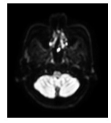
图 1 Ⅰ型,Ⅰa亚型。DWI:鼻咽黏膜显示欠清,尤其是鼻咽后壁黏膜呈明显低信号
Figure 1. Type I, Ia subtype. DWI:nasopharyngeal mucosa was unclear, an obvious low signal is observed in the posterior wall of the nasopharyngeal mucosa

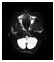
图 2 Ⅰ型,Ⅰb亚型。DWI:鼻咽黏膜不厚,双侧咽隐窝处局部信号对称性增高,接近于脊髓信号
Figure 2. Type I, Ib subtype. DWI:nasopharyngeal mucosa is not thick, bilateral pharyngeal recess at the local symmetry of the signals is increased close to the spinal cord signal

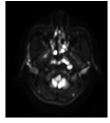
图 3 Ⅱ型。DWI:双侧鼻咽黏膜不对称,以咽隐窝处信号最高,接近于脊髓信号
Figure 3. Type Ⅱ。DWI:asymmetric bilateral nasopharyngeal mucosa, the pharyngeal recess is at the highest signal, close to the spinal cord signal

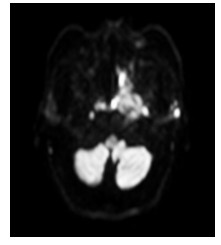
图 4 Ⅲ型,Ⅲa亚型。DWI:鼻咽黏膜双侧对称显影,未见明显增厚,以咽隐窝处信号最高,高于脊髓信号
Figure 4. Type Ⅲ, Ⅲa subtype. DWI: nasopharyngeal mucosa bilaterally symmetrical development; no obvious thickening; pharyngeal recess at the highest signal, higher than that of spinal cord signal

图 5 Ⅲ型,Ⅲb亚型。DWI:鼻咽黏膜对称性增厚,双侧咽隐窝处信号最高,高于脊髓信号
Figure 5. Type Ⅲ, Ⅲb subtype. DWI: nasopharyngeal mucosa thickening of bilateral symmetry; pharyngeal recess at the highest signal; signal is higher than that of the spinal cord

图 6 Ⅳ型,Ⅳa亚型。DWI:鼻咽左侧咽隐窝处黏膜结节状增厚,信号高于脊髓信号,左侧咽后淋巴结转移
Figure 6. Type Ⅳ, Ⅳa subtype. DWI: left side pharyngeal recess; nasopharyngeal mucosal nodular thickening; signal is higher than the spinal cord signal. Metastasis of left retro-pharyngeal lymph node is observed

图 7 Ⅳ型,Ⅳb亚型。DWI:鼻咽左侧壁、顶后壁黏膜明显增厚形成肿块灶,信号不均匀,最高信号高于脊髓信号
Figure 7. Type Ⅳ, Ⅳb subtype. DWI: left side wall; thickening observed in the top wall of the nasopharyngeal mucosa mass range with signal heterogeneity. The highest signal is higher than that of the spinal cord
表 1 DWI鼻咽黏膜分型与定性结果比较
Table 1. Comparison of DWI nasopharyngeal mucosa typing and qualitative results

-
[1] 祁建军, 栾阳, 高向东, 等.磁共振DWI和PWI在超急性脑梗死诊治中的应用[J].实用医学影像杂志, 2012, 13(22):69-71. http://www.cqvip.com/qk/84078x/201202/41643246.htmlQi JJ, Luan Y, Gao XD, et al. Application of MR DWI and PWI for emergent diagnosis and therapy of hyperacute cerebral infarction[J]. JPMI, 2012, 13(22):69-71. http://www.cqvip.com/qk/84078x/201202/41643246.html [2] Kitajima K, Yamasaki E, Kaji Y, et al. Comparison of DWI and PET/CT in evaluation of lymph node metastasis in uterine cancer [J]. World J Radiol, 2012, 4(5):207-214. https://www.ncbi.nlm.nih.gov/pmc/articles/PMC3386532/ [3] 陈玉芳, 程红岩.DWI在肝癌TACE后疗效评价的研究进展[J].中国医学计算机成像杂志, 2011, 17(5): 451-455. http://d.wanfangdata.com.cn/Periodical_zgyxjsjcx201105015.aspxChen YF, Cheng HY. Progress of DWI in evaluation of the potency of TACE for hepatocellular carcinoma[J]. Chin Comput Med Imag, 2011, 17(5):451-455. http://d.wanfangdata.com.cn/Periodical_zgyxjsjcx201105015.aspx [4] 王健, 姚忠秀, 饶圣祥, 等.3.0T磁共振弥散加权成像在胰腺癌中的应用价值[J].医学影像学杂志, 2012, 22(1):91-93. http://d.wanfangdata.com.cn/Periodical/yxyxxzz201201031Wang J, Yao ZX, Rao SX, et al. The applied value of breath-holding diffusion-weighter MR imaging for pancreatic carcinoma at 3.0T[J]. J Med Imaging, 2012, 22(1):91-93. http://d.wanfangdata.com.cn/Periodical/yxyxxzz201201031 [5] 郭立, 杨达宽, 袁曙光, 等.胰腺癌磁共振弥散成像中b值的选择[J].中国临床医学影像杂志, 2010, 21(2): 87-89. http://www.cqvip.com/Main/Detail.aspx?id=33011796Guo L, Yang DK, Yuan SS, et al. The best b value of MR diffusion-weighted imaging for pancreatic cancer[J]. J Chin Clin Med Imaging, 2010, 21(2):87-89. http://www.cqvip.com/Main/Detail.aspx?id=33011796 [6] 李琼, 白人驹, 孙浩然.MR DWI在淋巴瘤检出、疗效监测中的应用[J].中国医学影像技术, 2010, 26(12): 2313-2316. http://www.cnki.com.cn/Article/CJFDTotal-ZYXX201012039.htmLi Q, Bai RJ, Sun HR. Application of MR DWI in detection and therapeutic monitor of lymphoma[J]. Chin J Med Imaging Technol, 2010, 26(12):2313-2316. http://www.cnki.com.cn/Article/CJFDTotal-ZYXX201012039.htm [7] 张洪涛, 刘倩, 陆虹, 等.WB-DWI在乳腺癌骨转移诊断中的应用[J].临床放射学杂志, 2011, 30(9):1345-1348. http://www.cnki.com.cn/Article/CJFDTotal-LCFS201109030.htmZhang HT, Liu Q, Lu H, et al. Application of whole body diffusion weighted imaging in the diagnosis of the metastatic bone in breast cancer[J]. J Clin Radiol, 2011, 30(9):1345-1348. http://www.cnki.com.cn/Article/CJFDTotal-LCFS201109030.htm [8] 林建华, 谭理连, 何伟红, 等.全身弥散加权成像与同位素骨扫描诊断乳腺癌骨转移对比研究[J].放射学实践, 2011, 26(10):1107-1109. http://med.wanfangdata.com.cn/viewHTML/PeriodicalPaper_fsxsj201110023.aspxLin JH, Tan LL, He WH, et al. Comparison of the clinic value of whole body MR diffusion weighted imaging and radionuclide bone scan in the diagnosis of skeletal metastasis of breast cancer[J]. Radiol Practice, 2011, 26 (10):1107-1109. http://med.wanfangdata.com.cn/viewHTML/PeriodicalPaper_fsxsj201110023.aspx [9] 徐贤, 马林, 安宁豫, 等.全身磁共振弥散加权成像在鉴别恶性肿瘤骨转移和感染性病变中的应用[J].中国临床保健杂志, 2011, 14(2):120-122. http://www.cqvip.com/QK/71135X/201107/37730939.htmlXu X, Ma L, An NY, et al. Comparison on spinal infection and malignant tumor metastasis with whole body diffusion-weighted imaging[J]. Chin J Clin Health, 2011, 14(2):120-122. http://www.cqvip.com/QK/71135X/201107/37730939.html [10] 徐光炎, 金琼英, 沈巨峰, 等.MRI动态增强联合DWI对乳腺良恶性病变的鉴别[J].中国中西医结合外科杂志, 2012, 18(2):130-133. http://www.cqvip.com/QK/90865X/201301/45650026.htmlXu GY, Jin QY, Shen JF, et al. Dynamic contrast-enhanced MRI combined diffusion-weighted image on identification of benign and malignant breast lesions[J]. Chin J Surg Integrat Tradition Western Med, 2012, 18(2):130-133. http://www.cqvip.com/QK/90865X/201301/45650026.html [11] 刘彪.扩散加权成像与动态增强技术在乳腺病变诊断中的应用[J].临床放射学杂志, 2011, 30(12):1853-1856. http://www.cnki.com.cn/Article/CJFDTotal-LCFS201112038.htmLiu B. Value of DWI and DCE-MRI in diagnosis of breast lesions [J]. J Clin Radiol, 2011, 30(12):1853-1856. http://www.cnki.com.cn/Article/CJFDTotal-LCFS201112038.htm [12] 李卉, 谢传淼, 刘学文, 等.全身弥散加权成像在鼻咽癌患者诊断中的应用研究[J].中国肿瘤临床, 2011, 38(12):723-726. http://www.cjco.cn/cn/article/doi/doi:10.3969/j.issn.1000-8179.2011.12.009Li H, Xie CM, Liu XW, et al. The clinical application of whole-body diffusion weighted imaging of nasopharyngeal cancer [J]. Chin J Clin Oncol, 2011, 38(12):723-726. http://www.cjco.cn/cn/article/doi/doi:10.3969/j.issn.1000-8179.2011.12.009 [13] 李伟, 卢斌贵, 傅文海, 等.多层螺旋CT在鼻咽癌预后随访中的价值[J].广东医学, 2012, 33(2):231-233. http://d.wanfangdata.com.cn/Periodical/zgyyzn201315423Li W, Lu BG, Fu WH, et al. The value of multi-slice spiral CT in follow-up of nasopharyngeal carcinoma[J]. Guangdong Med J, 2012, 33(2):231-233. http://d.wanfangdata.com.cn/Periodical/zgyyzn201315423 [14] 杨百华, 陈英, 徐鹭英, 等.磁共振扩散加权成像在鉴别鼻咽癌放疗后鼻咽坏死与复发中的价值[J].中国癌症防治杂志, 2011, 3(4):298-302. http://cpfd.cnki.com.cn/Article/CPFDTOTAL-ZHYX201110001176.htmYang BH, Chen Y, Xu LY, et al. Values of diffusion- weighted imaging in differential diagnosing necrosis and recurrence of nasopharyngeal carcinoma after radiotherapy[J]. Chin J Oncol Prev Treat, 2011, 3(4):298-302. http://cpfd.cnki.com.cn/Article/CPFDTOTAL-ZHYX201110001176.htm [15] 韩晶, 刘念龙, 叶峰, 等.MRI扩散加权成像在鼻咽癌调强放疗疗效评价中的应用价值[J].医学影像学杂志, 2012, 22(10):1635-1638. http://d.wanfangdata.com.cn/Periodical/yxyxxzz201210014Han J, Liu NL, Ye F, et al. DWI-MRI in evaluating intensity modulated radiation therapy for nasopharyngeal carcinoma[J]. J Med Imaging, 2012, 22(10):1635-1638. http://d.wanfangdata.com.cn/Periodical/yxyxxzz201210014 [16] 龚晓昌, 李金高, 敖帆, 等.磁共振弥散成像在鼻咽癌复发诊断中的临床意义[J].实用癌症杂志, 2011, 26(4):395-397. http://www.cqvip.com/qk/95841x/201102/38785044.htmlGong XC, Li JG, Ao F, et al. The value of the diffusion-weighted MRI imaging in the diagnosis of relapse of nasopharyngeal carcinoma[J]. Practical J Cancer, 2011, 26(4):395-397. http://www.cqvip.com/qk/95841x/201102/38785044.html -

 下载:
下载:


 点击查看大图

计量
- 文章访问数: 37
- HTML全文浏览量: 10
- PDF下载量: 0
- 被引次数: 0
