
Automated breast volume imaging with color doppler ultrasound in evaluating effect of neoadjuvant chemotherapy for breast cancer
-
摘要:
目的 探讨自动乳腺全容积成像(ABVS)与彩色多普勒超声(CDFI)对乳腺癌新辅助化疗(NCT)疗效评价的意义。 方法 应用ABVS与CDFI对42例乳癌患者NCT前后的病灶成像特点进行观察与分析。 结果 42例乳癌患者接受NCT后, 其中完全缓解的患者ABVS从冠状面显示原发肿瘤分布范围及声像改变, CDFI图像显示原发灶内血流变化等均具有显著性差异(P < 0.01);而在无变化及部分缓解的病灶中的ABVS从冠状面显示原发肿瘤分布范围及声像改变, CDFI显示病灶内血流变化等均无显著性差异(P>0.05) 结论 利用ABVS与CDFI对乳癌NCT前后疗效评价具有较大的临床意义及价值。 Abstract:Objective To explore automatic breast full volume imaging (ABVS) and color doppler ultrasound (CDFI) for breast cancer neoadjuvant chemotherapy (NCT) in the evaluation of curative effect. Methods The application of a CDFI and ABVS 42 cases of breast cancer patients receiving NCT lesions imaging characteristics before and after observation and analysis. Results ABVS from coronal distribution and audio-visual change according to the primary tumor, and CDFI images showing the primary tumors and blood flow changes within all have significant difference in patients with complete remission after NCT (P < 0.01); ABVS from coronal distribution and audio-visual change according to the primary tumor, and CDFI showed lesions in blood flow changes, etc. in patients with the absence of change and easing some lesions have no significant difference (P>0.05). Conclusion We use ABVS and CDFI for breast cancer before and after the NCT curative effect evaluation of great clinical significance and value. -
Key words:
- ABVS /
- CDFI /
- breast cancer /
- neoadjuvant chemotherapy /
- therapeutic evaluation
-

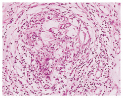
图 1 NCT前组织穿刺活检证实为乳腺浸润性导管癌(H&E×100)
Figure 1. Tissue biopsy confirmed invasive ductal carcinoma of the breast before NCT(H&E×100)

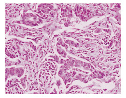
图 2 NCT后患者手术标本经病理学证实为G4级(H&E×100)
Figure 2. Surgical specimens of patients with histologically proven G4 level after NCT(H&E×100)

图 4 ABVS对该患者4个周期NCT后的冠状面成像
Figure 4. ABVS coronal imaging of the patient after four cycles of NCT
表 1 乳腺癌新辅助化疗疗效的临床评价与病理评价对比例
Table 1. Comparison of the clinical assessment and pathology evaluation of the neoadjuvant chemotherapy effect of breast cancer n

表 2 ABVS评价新辅助化疗前后肿瘤大小比较
Table 2. ABVS evaluation of tumor size before and after neoadjuvant chemotherapy

表 3 ABVS评价新辅助化疗前后肿瘤声像图的变化
Table 3. ABVS sonographic evaluation of tumor before and after neoadjuvant chemotherapy

表 4 CDFI在新辅助化疗前后对肿瘤血流类型变化的对比
Table 4. CDFI evaluation of blood flow of tumors before and after neoadjuvant chemotherapy

表 5 CDFI评价新辅助化疗前后肿瘤血流最高流速及阻力指数的变化
Table 5. CDFI evaluation of Vmax and RI in tumor before and after neoadjuvant chemotherapy

-
[1] Rastogi P. Anderson SJ, Bear HD, et al. Preoperative chemotherapy updates of National Surgical djuvant Breast and Bowel ProjectProtocolB-18 and B-27[J]. J Clin Oncol, 2008, 26(5):778-785. doi: 10.1200/JCO.2007.15.0235 [2] BearH D, Anderson S, SmithR E, et al. Sequential preoperative or postoperative docetaxel added to preoperative doxorubicin plus cyclophosphan ide for operable breast cancer National Surgical Adjuvant Breastand bowel Project Protocol B-27[J]. J Clin Oncol, 2006, 24(13):2019-2027. doi: 10.1200/JCO.2005.04.1665 [3] Tozaki M, Isobe S, Yamaguchi M, et al. Optimal scanning technique to cover the whole breast using an automated breast volume scanner[J]. Jpn J Radiol, 2010, 28(4):325-328. doi: 10.1007/s11604-010-0424-2 [4] Wojcinski S, Farrokh A. Hille U, et al. The automated breast volume scanner (ABVS):initial experiences in lesion detection compared with conventional handheld B-mode ultrasound:a pilot study of 50 cases[J]. Int J Womens Health, 2011, 3:337-346. http://www.ncbi.nlm.nih.gov/pubmed/22114526 [5] Lin X, Wang J, Han F, et al. Analysis of eighty-one cases with breast lesions using automated breast volume scanner and comparison with handheld ultrasound[J]. Eur J Radiol, 2012, 81(5):873. doi: 10.1016/j.ejrad.2011.02.038 [6] Kotsianos-hermle D, Hiltawsky KM, Wirth S, et al. Analysis of 107 breast lesions with automated 3D ultrasound and comparison with mammography and manual ultrasound[J]. Eur J Radiol, 2009, 71(1):109-115. doi: 10.1016/j.ejrad.2008.04.001 [7] Adler DD, Carson PL, Rubin JM, et al. Doppler ultrasound color flow imaging in the study of breast cancer:preliminary findings[J]. Ultrasound Med Biol, 1990, 16(6):553-559. doi: 10.1016/0301-5629(90)90020-D [8] Therasse P, Arbuck SG, Eisenhauer EA, et al. New guidelines to evaluate the response to treatment in solid tumors.European organization for reseach and treatment of cancer, national cancer institute of the unitedstates, National Cancer Institute of Canada[J]. J Natl Cancer Inst, 2000, 92(4):205-216. doi: 10.1056/NEJMoa1200694 [9] Smith IC, Hey SD, Hutcheon AW, et al. Neoadjuvant chemotherapy in breast cancer Significantly enhanced response with docetaxel [J]. J Clin Oncol, 2002, 20(6):1456. doi: 10.1200/JCO.2002.20.6.1456 [10] 李云凌, 赵斌, 冯鑫至.乳腺摄影与超声弹性成像对乳腺疾病诊断的对比性研究[J].医学影像学杂志, 2010, 20(10):1452-1455. http://d.wanfangdata.com.cn/Thesis_Y1822228.aspxLi Yunling, Zhao Bin, Feng Xin to the magazine. A comparative study on[J]. medical imaging, mammography and ultrasound elasticity imaging in the diagnosis of breast diseases, 2010, 20(10): 1452-1455. http://d.wanfangdata.com.cn/Thesis_Y1822228.aspx [11] Jos A, vanderHage, Comelis JH, et al. Preoperative chemotherapy in primary operable breast cancer results from the european organization for research and treatment of cancer trial[J]. J Clin Oncol, 2001, 19:4224-4237. doi: 10.1200/JCO.2001.19.22.4224 [12] Londero V, Bazzocchi M, Del Frate C, et al. Locally advanced breast cancer:comparison of mammography sonography and MR imagine in evaluation of residual disease in women receiving neoadjuvant Chemotherapy[J]. Eur Radiol, 2004, 14(8):1371-1379. doi: 10.1007/s00330-004-2246-z [13] 刘志聪, 滕淑琴, 蔡洁, 等.三维超声成像在乳腺疾病中的临床应用研究[J].中国超声诊断杂志, 2003, 4(10):751-753. http://www.cnki.com.cn/Article/CJFDTotal-CSZD200310013.htmLiu Zhicong, Teng Shuqin, Cai Jie, et al. Study on theclinical application of[J]. 3D ultrasound imaging in breast disease Chinese Journal of ultrasound diagnosis, 2003, 4(10):751-753. http://www.cnki.com.cn/Article/CJFDTotal-CSZD200310013.htm [14] 朱庆莉, 姜玉新, 孙强, 等.乳腺癌的彩色多普勒血流分布特征与组织病理学对照研究[J].中国医学影像技术, 2005, 21(10):1516-1518. http://www.cqvip.com/QK/98028X/200510/20321458.html Zhu Qingli, Jiang Yuxin, sun Qiang, et al. Breast cancercolor Doppler flow distribution characteristics andhistopathological study. Chinese medical imaging technology, 2005, 21(10):1516-1518. http://www.cqvip.com/QK/98028X/200510/20321458.html [15] Lee SW, Choi HY, Baek SY, et al. Role of color and power doppler imaging in differentiating between malignant and benign solid breast masses[J]. J Clin Ultrasound, 2002, 30(8):459-464. doi: 10.1002/jcu.10100 -

 下载:
下载:


 点击查看大图
计量
- 文章访问数: 45
- HTML全文浏览量: 9
- PDF下载量: 3
- 被引次数: 0
