
Influence of tumor microenvironment on culture and identification of breast cancer stem cell-like microspheres
-
摘要:
目的 探讨肿瘤微环境在乳腺癌干细胞(breast cancer stem cells,BCSCs)培养鉴定及分化过程中的影响及意义。方法:采用无血清培养液PCM-2及成纤维细胞上清液对乳腺癌细胞及MCF-7细胞进行原代培养。观察乳腺癌细胞微球体形成状况,MTT比色法检测乳腺癌细胞的增殖能力,免疫细胞化学 方法 检测乳腺癌干细胞标记物及上皮间质标记物的表达,并通过RT-PCR进行验证。 结果 无血清培养液PCM-2培养的原代细胞微球体的直径大于成纤维细胞上清液的培养(t=4.996,P= 0.002),且原代细胞中ALDH1(aldehyde dehydrogenase 1)的表达率高于后者。成纤维细胞上清液培养的细胞生长速度较无血清培养液PCM-2快,差异具有统计学意义(P=0.004)。RT-PCR检测发现无血清培养液PCM-2培养的原代细胞中ALDH1表达上调,E-cadherin、Vimentin表达下调。 结论 在乳腺癌原代细胞和MCF-7细胞中可以采用无血清悬浮培养方法富集BCSCs样微球体,成纤维细胞上清液能够促进BCSCs样微球体的增殖与分化。提示乳腺肿瘤微环境在乳腺癌细胞的生长增殖过程中发挥了至关重要的作用。 Abstract:Objective To investigate the influence and significance of tumor microenvironment in breast cancer stem-cell culture and identification. Methods Cells isolated from primary breast cancer tissues were cultured in vitro in a serum-free medium PCM-2 and in the supernatant of cultured fibroblasts. The MCF-7 breast cancer cell line was used as the control group. The status of the microspheres was observed, and the proliferative capacity of the cells was detected by methyl thiazolyl tetrazolium assay. The expression of stem cell and epithelial-mesenchymal markers were detected by real-time reverse transcription polymerase chain reaction. Results The diameter of microspheres in PCM-2 gradually increased with prolonged incubation time (t=4.996, P=0.002). The cells in the supernatant of cultured fibroblasts increased daily and mostly exhibited a spindle cell growth. The growth rate of primary breast cancer cells was faster in the supernatant of cultured fibroblasts than in PCM-2 (P=0.004). Compared with the case of cells in the supernatant of cultured fibroblasts, aldehyde dehydrogenase 1 was upregulated in the primary breast cancer cells cultured in serum-free medium PCM-2, whereas E-cadherin and Vimentin were downregulated. Conclusion Serum-free culture can be one of the best methods for enriching breast cancer stem cell-like mammospheres. The tumor micro-environment serves a vital function in the growth and development of tumor cells and in the evolution of breast cancer. -
Key words:
- breast cancer /
- breast cancer stem cells /
- tumor microenvironment /
- serum-free culture /
- mammospheres
-

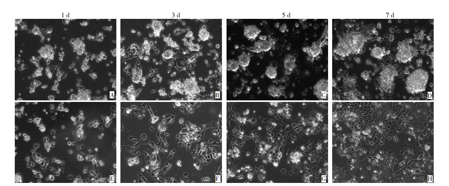
图 1 无血清培养液PCM-2及成纤维细胞上清液培养的原代细胞微球体的形成状况(倒置相差显微镜×200)
Figure 1. Status of microspheres formed in two different culture mediums(IPCM×200)
A-D: Serum-free medium PCM-2; E-H: Supernatant of cultured fibroblasts

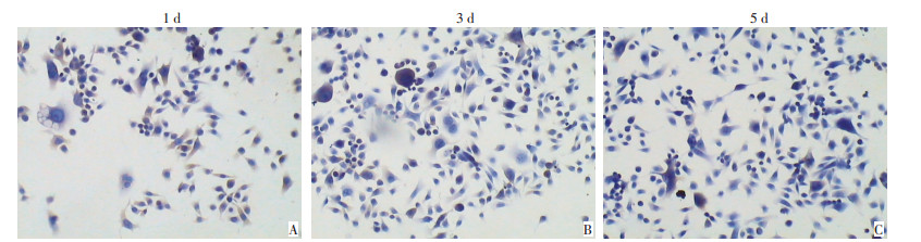
图 2 无血清培养液PCM-2培养的原代细胞中ALDH1表达(SP×200)
Figure 2. Expression of ALDH1 in primary breast cancer cells(SP × 200)
A: Expression of ALDH1 in 1d; B: Expression of ALDH1 in 3d; C: Expression of ALDH1 in 5d

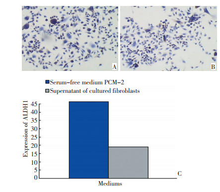
图 3 无血清培养液PCM-2及成纤维细胞上清液培养的原代细胞中ALDH1表达(SP×200)
Figure 3. Expression of ALDH1 in primary breast cancer cells in PCM-2 and the supernatant of cultured fibroblasts(SP × 200)
A:Serum-free medium PCM-2; B:Supernatant of cultured fibroblasts; C: Expression of ALDH1 in primary breast cancer cells in two different mediums

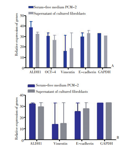
图 4 无血清培养液PCM-2及成纤维细胞上清液培养的乳腺癌细胞第3天时干细胞及上皮间质标记物表达
Figure 4. Expression of stem cells and epithelial-mesenchymal markers in MCF-7 and primary breast cancer cells cultured in PCM-2 and in the supernatant of cultured fibroblasts for 3 d
A: Expression of different genes in MCF-7; B: Expression of different genes in primary breast cancer cells
表 1 RT-PCR相关引物序列表
Table 1. The sequence of relative primers in real-time reverse transcrip⁃ tion polymerase chain reaction

-
[1] Ferlay J, Shin HR, Bray F, et al. Estimates of worldwide burden of cancer in 2008: GLOBOCAN 2008[J]. Int J Cancer, 2010, 127(12):2893-2917. doi: 10.1002/ijc.25516 [2] Al-Hajj M, Wicha MS, Benito-Hernandez A, et al. Prospective identification of tumorigenic breast cancer cells[J]. Proc Natl Acad Sci U S A, 2003, 100(7):3983-3988. doi: 10.1073/pnas.0530291100 [3] Ginestier C, Hur MH, Charafe-Jauffret E, et al. ALDH1 is a marker of normal and malignant human mammary stem cells and a predictor of poor clinical outcome[J]. Cell Stem Cell, 2007, 1(5):555-567. doi: 10.1016/j.stem.2007.08.014 [4] Nagy JA, Chang SH, Shih SC, et al. Heterogeneity of the tumor vasculature[J]. Semin Thromb Hemost, 2010, 36(3):321-331. [5] Mantel PY, Schmidt-Weber CB. Transforming growth factor-beta: recent advances on its role in immune tolerance[J]. Methods Mol Biol, 2011, 677:303-338. [6] Kalluri R, Weinberg RA. The basics of epithelial-mesenchymal transition[J]. J Clin Invest, 2009, 119(6):1420-1428. doi: 10.1172/JCI39104 [7] Lengerke C, Fehm T, Kurth R, et al. Expression of the embryonic stem cell marker SOX2 in early-stage breast carcinoma[J]. BMC Cancer, 2011, 11:42. doi: 10.1186/1471-2407-11-42 [8] Debeb BG, Xu W, Mok H, et al. Differential radiosensitizing effect of valproic acid in differentiation versus self-renewal promoting culture conditions[J]. Int J Radiat Oncol Biol Phys, 2010, 76(3):889-895. doi: 10.1016/j.ijrobp.2009.09.052 [9] Ricardo S, Vieira AF, Gerhard R, et al. Breast cancer stem cell markers CD44, CD24 and ALDH1: expression distribution within intrinsic molecular subtype[J]. J Clin Pathol, 2011, 64(11):937-946. doi: 10.1136/jcp.2011.090456 [10] Xing F, Saidou J, Watabe K. Cancer associated fibroblasts (CAFs) in tumor microenvironment[J]. Front Biosci (Landmark Ed), 2010, 15:166-179. doi: 10.2741/3613 [11] Sneddon JB, Zhen HH, Montgomery K, et al. Bone morphogenetic protein antagonist gremlin 1 is widely expressed by cancer-associated stromal cells and can promote tumor cell proliferation[J]. Proc Natl Acad Sci U S A, 2006, 103(40):14842-14847. doi: 10.1073/pnas.0606857103 [12] Römer AM, Lühr I, Klein A, et al. Normal mammary fibroblasts induce reversion of the malignant phenotype in human primary breast cancer[J]. Anticancer Res, 2013, 33(4):1525-1536. http://www.wanfangdata.com.cn/details/detail.do?_type=perio&id=9cb45ac9ef3bb5b1362f9a86ba87c5c1 [13] Polyak K, Kalluri R. The role of the microenvironment in mammary gland development and cancer[J]. Cold Spring Harb Perspect Biol, 2010, 2(11):a003244. [14] Korkaya H, Liu S, Wicha MS. Breast cancer stem cells, cytokine networks, and the tumor microenvironment[J]. J Clin Invest, 2011, 121 (10):3804-3809. doi: 10.1172/JCI57099 [15] Orimo A, Gupta PB, Sgroi DC, et al. Stromal fibroblasts present in invasive human breast carcinomas promote tumor growth and angiogenesis through elevated SDF-1/CXCL12 secretion[J]. Cell, 2005, 121(3):335-348. doi: 10.1016/j.cell.2005.02.034 -

 下载:
下载:
 点击查看大图
计量
- 文章访问数: 84
- HTML全文浏览量: 4
- PDF下载量: 1
- 被引次数: 0
