
Correlation and clinical significance between the breast cancer MRI schedule of reinforcement and the pattern of tumorshrinkage after neo-adjuvant chemotherapy
-
摘要:
目的 探讨乳腺癌的磁共振强化方式与新辅助化疗后退缩模式间的相关性,及其指导制定手术方案的临床意义。 方法 55例局部进展期乳腺癌患者分别于新辅助化疗前及全程化疗后手术前,行乳腺磁共振动态增强扫描,观察化疗前肿瘤的磁共振强化方式和化疗后肿瘤退缩模式,分析两者之间的相关性。 结果 55例患者中54例为单乳病变、1例为双乳病变,共56处病灶。肿块样强化为24处(43%),其中23处呈向心性退缩、1处呈环形退缩(P < 0.01);多灶肿块样强化为13处(23%),其中2处为单一肿块呈向心性退缩、11处退缩后仍呈多灶肿块(P < 0.01);肿块伴周边非肿块样强化为8处(14%),其中4处呈向心性退缩、4处呈蜂窝状多灶退缩(P>0.05);非肿块样强化为11处(20%),其中4处呈向心性退缩、7处呈蜂窝状多灶退缩(P < 0.01)。 结论 新辅助化疗后肿瘤的退缩模式是保乳手术选择的关键因素之一,根据乳腺癌在新辅助化疗前的磁共振动态增强强化方式,可在一定程度上预测其在化疗后的退缩模式,进而预测患者保乳手术的可行性,为临床医生选择个体化的后续治疗方案提高患者生存质量提供参考。 Abstract:Objective To investigate the correlation between the breast cancer MRI schedule of reinforcement and the shrinkage pattern of tumor after neo-adjuvant chemotherapy, and its clinical significance in the guidance of formulating operation plan. Methods Dynamic contrast-enhanced MRI scan was performed before chemotherapy and before surgery after a whole-range N-Acety-L-Cysteine (NAC) treatment in 55 patients with loco-regionally advanced breast cancer who received the neo-adjuvant chemotherapy. MRI schedule of reinforcement and the shrinkage pattern of tumor after neo-adjuvant chemotherapy were obtained in the treatment, and the correlation between the two was analyzed. Results Of the 55 patients, the unilateral breast mass was found in 54 and the bilateral lesion in 1. There were 56 neo-plastic foci in these patients. The mass-like enhanced image was seen in 24 of the total cases (43%), of which 23 presented with a centripetal shrinkage, 1 with an annular decline, (P < 0.01). Multifocal mass-like enhancement image was found in 13 of the total cases, (23%), of which 2 centripetal shrinkages were the singular mass, 11 remained a multifocal lesion after the tumor shrinkage (P < 0.01). The mass with peripheral non-tumor-like enhancement image was seen in 8 of the total cases (14%), of which 4 showed a centripetal shrinkage and another 4 a honeycombed multifocal decline (P>0.05). There were 11 of the total cases with non-tumor-like enhancement (20%), in which 4 assumed a centripetal shrinkage and 7 a honeycombed multifocal shrinkage (P < 0.01). Conclusion The tumor shrinking pattern and its accurate radiological image evaluation are the keys to the selection of breast-conserving surgery and the control of local recurrence after treatment of NAC regimen. We can predict the shrinking pattern through the type of the lesion on baseline before NAC, which is important for the patients and surgeon to get a reasonable expectation in the subsequent treatments. -

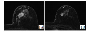
图 1 浸润性导管癌向心性退缩的MRI动态增强图像
Figure 1. Dynamic contrast-enhanced MRI of centripetal shrinkage pattern in invasive ductal carcinoma
1A: A mass-like enhancement of primary neo-plastic foci before neo-adjuvant chemotherapy; 1B: A centripetal shrinkage of the neo-plastic foci after the chemotherapy

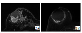
图 2 化生性癌环形退缩的MRI动态增强图像
Figure 2. Dynamic contrast-enhanced MRI image of annular shrinkage pattern in metaplastic carcinoma
2A: Before chemotherapy, the shape of the primary tumor on MRI was cystic-sold; 2B: Annular shrinkage of tumor after the chemotherapy

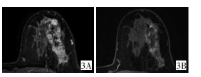
图 3 浸润性导管癌蜂窝状退缩的MRI动态增强图像
Figure 3. Dynamic contrast-enhanced MRI of honeycombed shrinkage pattern in invasive ductal carcinoma
3A: The shape of the primary tumor on MRI was diffuse before chemotherapy; 3B: Honeycombed shrinkage of the tumor after the chemotherapy
表 1 新辅助化疗前56处病灶磁共振强化方式与化疗后退缩模式比较
Table 1. Comparison of MRI enhancement mode before neo-adjuvant chemotherapy and with the pattern of tumor shrinkage after the chemotherapy in 56 breast cancer cases undergoing NAC regimen

-
[1] Sweeting RS, Klauber-Demore N, Meyers MO, et al. Young women with locally advanced breast cancer who achieve breast conservation after neoadjuvant chemotherapy have a low local recurrence rate[J]. Am Surg, 2011, 77(7):880-855. [2] 周娟, 邢旭东, 李功杰, 等.动态增强磁共振成像对新辅助化疗后乳腺癌残余病灶的术前评估[J].实用放射学杂志, 2012, 28(10):1553-1556. doi: 10.3969/j.issn.1002-1671.2012.10.017Zhou J, Xing XD, Li GJ, et al. Preoperative evaluation of residual breast carcinoma by dynamic contrast-enhanced MR imaging after neoadjuvant chemotherapy[J]. J Pract Radiol, 2012, 28(10):1553-1556. doi: 10.3969/j.issn.1002-1671.2012.10.017 [3] 胡静, 赵亚娥, 汪登斌, 等. MRI对侵润性乳腺癌新辅助化疗疗效的评估价值[J].外科理论与实践, 2011, 16(1):29-34.Hu J, Zhao YE, Wang DB, et al. Evaluation of the therapeutic effects of neoadjuvant chemotherapy in invasive breast cancer using magnetic resonance imaging[J]. J Surg Concepts Pract, 2011, 16(1): 29-34. [4] Lobbes MB, Prevos R, Smidt M, et al. The role of magnetic resonance imaging in assessing residual disease and pathologic complete response in breast cancer patients receiving neoadjuvant chemotherapy: a systematic review[J]. Insights Imaging, 2013, 4(2):163-175. doi: 10.1007/s13244-013-0219-y [5] Straver ME, Loo CE, Rutgers EJ, et al. MRI-model to guide the surgical treatment in breast cancer patients after neoadjuvant chemotherapy[J]. Ann Surg, 2010, 251(4):701-707. doi: 10.1097/SLA.0b013e3181c5dda3 [6] Bahri S, Chen JH, Mehta RS, et al. Residual breast cancer diagnosed by MRI in patients receiving neoadjuvant chemotherapy with and without bevacizumab[J]. Ann Surg Oncol, 2009, 16(6):1619-1628. doi: 10.1245/s10434-009-0441-5 [7] Kim HJ, Im YH, Han BK, et al. Accuracy of MRI for estimating residual tumor size after neoadjuvant chemotherapy in locally advanced breast cancer: relation to response patterns on MRI[J]. Acta Oncol, 2007, 46(7):996-1003. doi: 10.1080/02841860701373587 [8] Peintinger F, Kuerer HM, McGuire SE, et al. Residual specimen cellularity after neoadjuvant chemotherapy for breast cancer[J]. Br J Surg, 2008, 95(4):433-437. http://www.wanfangdata.com.cn/details/detail.do?_type=perio&id=10.1002/bjs.6044 [9] Loo CE, Teertstra HJ, Rodenhuis S, et al. Dynamic contrast-enhanced MRI for prediction of breast cancer response to neoadjuvant chemotherapy: initial results[J]. AJR Am J Roentgenol, 2008, 191(5):1331-1338. doi: 10.2214/AJR.07.3567 [10] Akazawa K, Tamaki Y, Taguchi T, et al. Preoperative evaluation of residual tumor extent by three-dimensional magnetic resonance imaging in breast cancer patients treated with neoadjuvant chemotherapy[J]. Breast J, 2006, 12(2):130-137. http://www.wanfangdata.com.cn/details/detail.do?_type=perio&id=731af2c6cf36923a3dc902bbe7e58393 [11] Rosen EL, Blackwell KL, Baker JA, et al. Accuracy of MRI in the detection of residual breast cancer after neoadjuvant chemotherapy[J]. AJR Am J Roentgenol, 2003, 181(5):1275-1282. doi: 10.2214/ajr.181.5.1811275 [12] 尹波, 刘莉, 张碧云, 等. MRI在乳腺癌新辅助化疗残留病灶评价中的价值[J].实用放射学杂志, 2013, 29(9):1441-1444. doi: 10.3969/j.issn.1002-1671.2013.09.016Yin B, Liu L, Zhang BY, et al. The value of MRI in evaluation of residual tumor to neoadjuvant chemotherapy for breast cancer[J]. J Pract Radiol, 2013, 29(9):1441-1444. doi: 10.3969/j.issn.1002-1671.2013.09.016 [13] Garimella V, Qutob O, Fox JN, et al. Recurrence rates after CE-MRI image guided planning for breast-conserving surgery following neoadjuvant chemotherapy for locally advanced breast cancer patients[J]. Eur J Surg Oncol, 2007, 33(2):157-161. doi: 10.1016/j.ejso.2006.09.019 [14] Loo CE, Straver ME, Rodenhuis S, et al. Magnetic resonance imaging response monitoring of breast cancer during neoadjuvant chemotherapy: relevance of breast cancer subtype[J]. J Clin Oncol, 2011, 29(6):660-666. doi: 10.1200/JCO.2010.31.1258 [15] Hayashi Y, Takei H, Nozu S, et al. Analysis of complete response by MRI following neoadjuvant chemotherapy predicts pathological tumor responses differently for molecular subtype of breast cancer[J]. Oncol Lett, 2013, 5(1):83-89. doi: 10.3892/ol.2012.1004 -

 下载:
下载:
 点击查看大图
计量
- 文章访问数: 23
- HTML全文浏览量: 17
- PDF下载量: 0
- 被引次数: 0
