
Clinical significance of expression and phosphorylation of FAK in human osteosarcoma
-
摘要:
目的 探讨骨肉瘤组织中黏着斑激酶(focal adhesion kinase,FAK)的表达情况及其与临床病理特征和预后的关系。 方法 收集1999年12月至2011年l2月东南大学附属中大医院和南京军区南京总医院收治的经手术病理确诊的骨肉瘤标本113例,免疫组织化学检测病灶中FAK表达量及磷酸化水平,并通过RNA干扰观察改变FAK的表达与激活水平对骨肉瘤细胞增殖、凋亡、迁移及侵袭活动的影响。 结果 70例标本(61.95%)FAK过表达,其中42例(37.17%)pFAK阳性。FAK表达谱与性别、年龄、AJCC ⅡA/ⅡB期、病灶部位、术式、术前化疗效果等临床病理参数均无显著相关性。生存分析显示FAK的过表达及其磷酸化显著缩短了总生存时间(overall survival,OS)和无转移生存时间(metastasis-free survival,mFS),其与术前化疗效果是预测骨肉瘤生存时间和转移早晚的两个独立预后因素。细胞学实验则显示FAK的表达和激活可以促进骨肉瘤细胞的增殖、迁移及侵袭活动,并能抑制凋亡。 结论 FAK的过表达及其磷酸化与骨肉瘤恶性程度密切相关,可为生存期及预后判断提供一定参考。 Abstract:Objective To examine expression patterns of focal adhesion kinase (FAK) and its activated form, phosphorylated FAK (pFAK), in human osteosarcoma and to investigate the correlation of FAK expression with clinicopathological parameters and prognosis. Functional consequence of manipulating FAK protein levels was also investigated in human osteosarcoma cell lines. Methods Immunohistochemical staining was used to detect FAK and pFAK levels in pathologically archived materials from 113 patients with primary osteosarcoma. KaplanMeier survival and Cox regression analyses were used to evaluate prognoses. The role of FAK in cytological behavior of MG63 and 143B human osteosarcoma cell lines was studied via the FAK protein knockdown with siRNA. Cell proliferation, migration, invasiveness, and apoptosis were assessed using cell counting kit-8, Transwell, and Annexin V/PI staining methods. Results Both FAK and pFAK were overexpressed in osteosarcoma patients. Tumor cells exhibited cytoplasmicity and occasional membranous immunoreactivity for FAK. A total of 42 cases (37.17%) mainly showed expressed pFAK in cytoplasm of osteosarcoma cells. No overexpression staining of anti-FAK and anti-pFAK antibodies was observed in normal cancellous bone tissues or negative controls. Significant differences were observed in overall survival between FAK-/pFAK-and FAK+/pFAK-groups (P=0.016), FAK+/pFAK-and FAK+/pFAK+ groups (P=0.012), and FAK-/pFAK-and FAK+/ pFAK+ groups (P < 0.001). All groups showed similar metastasis-free survival. Cox proportional hazard analysis showed that FAK expression profile is an independent indicator of both overall and metastasis-free survival. siRNA-based knockdown of FAK significantly reduced migration and invasion of MG63 and 143B cells and affected proliferation and apoptosis in osteosarcoma cells. Conclusion Osteosarcoma malignancies in vitro and in vivo were correlated with overexpression and phosphorylation of FAK. These findings suggest that FAK plays an important biological role in osteosarcoma carcinogenesis. This study provides a better understanding of diagnostic and prognostic relevance of FAK overexpression and phosphorylation in osteosarcoma patients. Therefore, FAK and pFAK can be used as independent predictors of overall and metastasis-free survival in osteosarcoma patients. -
Key words:
- osteosarcoma /
- focal adhesion kinase /
- prognosis /
- migration /
- invasion
-

图 1 骨肉瘤与正常松质骨组织的FAK免疫组织化学染色和磷酸化FAK染色
Figure 1. Immunohistochemical staining of FAK (A, B, E, and G) and pFAK (C, D, F, and H) proteins in osteosarcoma cells and normal cancellous bone tissues
A. FAK was overexpressed in 61.95% (70/113) of osteosarcoma cells. Tumor cells exhibited cytoplasmic and occasional membranous immunoreactivity for FAK. B. Low FAK immunohistochemical staining was detected in 43 osteosarcoma patients. C. pFAK was mainly expressed in cytoplasm of osteosarcoma cells in 37.17% (42/113) of cases. D. Low pFAK immunohistochemical staining was detected in 71 osteosarcoma patients. E. No FAK immunohistochemical staining was observed in normal cancellous bone tissues. F. No pFAK immunohistochemical staining was observed in normal cancellous bone tissues. G. FAK immunohistochemical staining was not observed in negative controls. H. pFAK immunohistochemical staining was not observed in negative controls. Results shown here are representative images with ×400 magnification

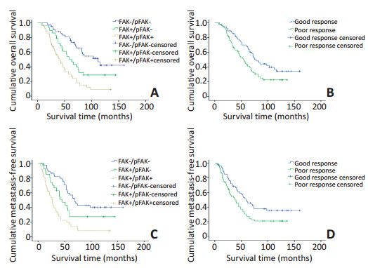
图 2 FAK表达水平及术前化疗效果不同的各组患者OS和mFS的Kaplan-Meier单因素分析
Figure 2. Kaplan-Meier analysis of overall and metastasis-free survival for patients with different FAK expression levels and different histological responses to pre-operative chemotherapy
A. Kaplan-Meier analysis of overall survival in osteosarcoma patients. The blue, green, and brown lines represent FAK-/pFAK-, FAK +/pFAK-, and FAK +/pFAK + groups, respectively. B. Kaplan-Meier analysis of overall survival in osteosarcoma patients. The blue line represents patients with good histological response to preoperative chemotherapy, and the green line represents patients with poor histological response to pre-operative chemotherapy. C. Kaplan-Meier analysis of metastasisfree survival in osteosarcoma patients. The blue green, and brown lines represent the FAK-/pFAK-, FAK+/pFAK-, and FAK+/pFAK+ groups, respectively. D. Kaplan-Meier analysis of metastasis-free survival in osteosarcoma patients. The blue line represents patients with good histological response to pre-operative chemotherapy, and the green line represents patients with poor histological response to pre-operative chemotherapy

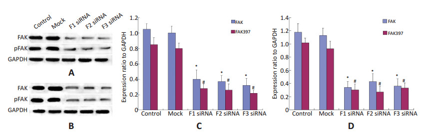
图 3 Western blot检测RNA干扰对于骨肉瘤细胞FAK蛋白表达及磷酸化水平的影响
Figure 3. Protein expression and phosphorylation of FAK in siRNA-treated osteosarcoma, control, and mock group cells
A. Western blot analysis of FAK and pFAK in MG-63 cells from control, mock, F1 siRNA, F2 siRNA, and F3 siRNA groups. GAPDH was used as control for protein load and integrity. B. Western blot analysis of FAK and pFAK in 143B cells from control, mock, F1 siRNA, F2 siRNA, and F3 siRNA groups. GAPDH was used as control for protein load and integrity. C. Bar chart revealing the ratio of FAK and pFAK protein to GAPDH by densitometry in MG-63 cells. Data are presented as means ± SEM. D. Bar chart showing ratio of FAK and pFAK protein to GAPDH by densitometry in 143B cells. Data are presented as means ± SEM.

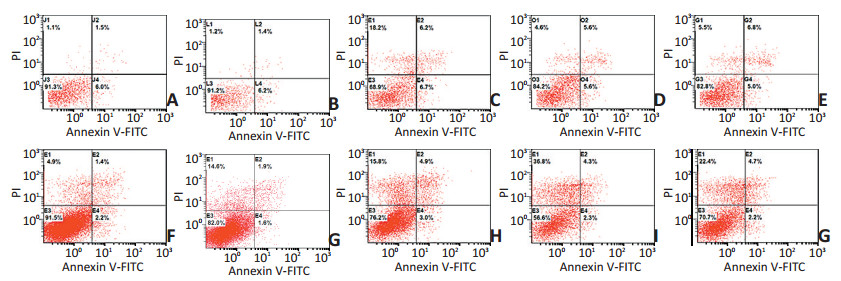
图 4 FAK表达对骨肉瘤细胞凋亡的影响
Figure 4. Effect of inhibiting FAK expression on apoptosis of osteosarcoma cells.
MG-63 cells treated with vehicle control (A), non-specific (scrambled) siRNA (B) or FAK siRNA of F1 (C), F2 (D), and F3 (E) were analyzed by flow cytometry after staining with annexin V-FITC/PI. 143B cells treated with vehicle control (F), non-specific (scrambled) siRNA (G), or FAK siRNA of F1 (H), F2 (I), and F3 (J) were analyzed by flow cytometry after staining with annexin V-FITC/PI

图 5 FAK表达对骨肉瘤细胞迁移能力的影响
Figure 5. Effect of inhibiting FAK expression on migration of osteosarcoma cells
A. Knockdown of FAK-inhibited cell migration by transwell assays. Penetration rate through the membrane was higher in non-transfected cells (control group) and non-specific (scrambled) siRNA transfected cells (mock group) than in FAK/RNAi cells (200×magnification); B. Number of cells that migrated to undersurface of the membrane as counted in six fields. Bars, SD. n=5, *P < 0.05

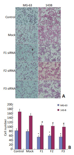
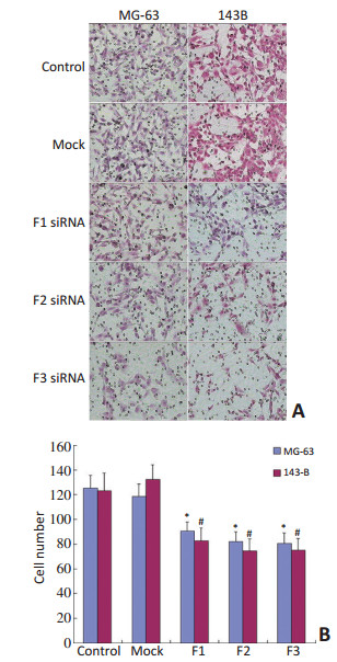
图 6 FAK表达对骨肉瘤细胞侵袭能力的影响
Figure 6. Effect of inhibiting FAK expression on invasion of osteosarcoma cells
A. Knockdown of FAK-inhibited cell invasion by transwell assays. Invasion ability of non-transfected cells (control group) and non-specific (scrambled) siRNA transfected cells (mock group) was higher than that of FAK/RNAi cells (200×magnification); B. Number of cells that passed through the membrane as counted in six fields. Bars, SD. n=5, *P < 0.05
表 1 AJCC分期Ⅱ期的骨肉瘤患者FAK表达谱与临床病理特征之间的关系
Table 1. Association of clinicopathological data and FAK expression profiles in patients with stage Ⅱ AJCC stage extremity osteosarcoma

表 2 骨肉瘤患者OS和mFS的Kaplan-Meier单因素分析
Table 2. Univariate analyses of factors associated with OS and MFS

表 3 骨肉瘤患者预后的多因素生存分析
Table 3. Multivariate analysis of factors associated with OS and MFS

表 4 FAK的siRNA转染对骨肉瘤细胞增殖的影响
Table 4. Effect of FAK siRNA transfection on cell proliferation

表 5 FAK的siRNA转染对骨肉瘤细胞凋亡的影响
Table 5. Effect of FAK siRNA transfection on cell apoptosis

-
[1] Hattinger CM, Serra M. Role of pharmacogenetics of drug-metabolizing enzymes in treating osteosarcoma[J]. Expert Opin Drug Metab Toxicol, 2015, 11(9):1449-1463. doi: 10.1517/17425255.2015.1060220 [2] He A, Qi W, Huang Y, et al. CD133 expression predicts lung metastasis and poor prognosis in osteosarcoma patients: A clinical and experimental study[J]. Exp Ther Med, 2012, 4(3):435-441. https://www.ncbi.nlm.nih.gov/pmc/articles/PMC3503795/ [3] Sun R, Shen J, Gao Y, et al. Overexpression of EZH2 is associated with the poor prognosis inosteosarcoma and function analysis indicates a therapeutic potential[J]. Oncotarget, 2016, 7(25):38333-38346. http://www.impactjournals.com/oncotarget/index.php?journal=oncotarget&page=article&op=view&path%5b%5d=9518 [4] Hattinger CM, Fanelli M, Tavanti E, et al. Advances in emerging drugs for osteosarcoma[J]. Expert Opin Emerg Drugs, 2015, 20(3): 495-514. doi: 10.1517/14728214.2015.1051965 [5] Lai IR, Chu PY, Lin HS, et al. Phosphorylation of focal adhesion kinase at Tyr397 in gastric carcinomas and its clinical significance[J]. Am J Pathol, 2010, 177(4):1629-1637. doi: 10.2353/ajpath.2010.100172 [6] Cheng YJ, Zhu ZX, Zhou JS, et al. Silencing profilin-1 inhibits gastric cancer progression via integrin β1/focal adhesion kinase pathway modulation[J]. World J Gastroenterol, 2015, 21(8):2323-2335. doi: 10.3748/wjg.v21.i8.2323 [7] Tzenaki N, Aivaliotis M, Papakonstanti EA. Focal adhesion kinasephosphorylates the phosphatase and tensin homolog deleted on chromosome 10 under the control of p110δ phosphoinositide-3 kinase[J]. FASEB J, 2015, 29(12):4840-4852. doi: 10.1096/fj.15-274589 [8] Thanapprapasr D, Previs RA, Hu W, et al. PTEN expression as a predictor of response to focal adhesion kinaseinhibition in uterine cancer[J]. Mol Cancer Ther, 2015, 14(6):1466-1475. doi: 10.1158/1535-7163.MCT-14-1077 [9] Zeng XQ, Li N, Ma LL, et al. Prognostic value of focal adhesion kinase (FAK) in human solid carcinomas: A Meta-analysis[J]. PLoS One, 2016, 11(9):e0162666. doi: 10.1371/journal.pone.0162666 [10] Golubovskaya VM, Conway-Dorsey K, Edmiston SN, et al. FAK overexpression and p53 mutations are highly correlated in human breast cancer[J]. Int J Cancer, 2009, 125(7):1735-1738. doi: 10.1002/ijc.v125:7 [11] Lark AL, Livasy CA, Dressler L, et al. High focal adhesion kinase expression in invasive breast carcinomas is associated with an aggressive phenotype[J]. Mod Pathol, 2005, 18(10):1289-1294. doi: 10.1038/modpathol.3800424 [12] Li F, Zhang X, Jin YP, et al. Antibody ligation of human leukocyte antigen lass I molecules stimulates migration and proliferation of smooth muscle cells in a focal adhesion kinase-dependent manner [J]. Hum Immunol, 2011, 72(12):1150-1159. doi: 10.1016/j.humimm.2011.09.004 [13] Li G, Du X, Vass WC, et al. Full activity of the deleted in liver cancer 1 (DLC1) tumor suppressor depends on an LD-like motif that binds talin and focal adhesion kinase (FAK)[J]. Proc Natl Acad Sci U S A, 2011, 108(41):17129-17134. doi: 10.1073/pnas.1112122108 [14] Eke I, Cordes N. Dual targeting of EGFR and focal adhesion kinase in 3D grown HNSCC cell cultures[J]. Radiother Oncol, 2011, 99(3):279-286. doi: 10.1016/j.radonc.2011.06.006 [15] Zhao J, Guan JL. Signal transduction by focal adhesion kinase in cancer[J]. Cancer Metastasis Rev, 2009, 28(1-2):35-49. doi: 10.1007/s10555-008-9165-4 [16] Gómez Del Pulgar T, Cebrián A, Fernández-Aceñero MJ, et al. Focal adhesion kinase: predictor of tumour response and risk factor for recurrence after neoadjuvant chemoradiation in rectal cancer[J]. J Cell Mol Med, 2016, 20(9):1729-1736. doi: 10.1111/jcmm.2016.20.issue-9 [17] Kong D, Chen F, Sima NI. Inhibition of focal adhesion kinase induces apoptosis in bladder cancer cells via Src and the phosphatidylinositol 3-kinase/Akt pathway[J]. Exp Ther Med, 2015, 10(5):1725-1731. https://www.ncbi.nlm.nih.gov/pmc/articles/PMC4665970/ [18] Aronsohn MS, Brown HM, Hauptman G, et al. Expression of focal adhesion kinase and phosphorylated focal adhesion kinase insquamous cell carcinoma of the larynx[J]. Laryngoscope, 2003, 113(11): 1944-1948. https://www.researchgate.net/publication/9021787_Expression_of_Focal_Adhesion_Kinase_and_Phosphorylated_Focal_Adhesion_Kinase_in_Squamous_Cell_Carcinoma_of_the_Larynx [19] Ding L, Sun X, You Y, et al. Expression of focal adhesion kinase and phosphorylated focal adhesion kinase in human gliomas is associated with unfavorable overall survival[J]. Transl Res, 2010, 156(1):45-52. doi: 10.1016/j.trsl.2010.05.001 [20] Kaushik S, Ravi A, Hameed FM, et al. Concerted modulation of paxillin dynamics at focal adhesions by deleted in liver cancer-1 and focal adhesion kinase during early cell spreading[J]. Cytoskeleton (Hoboken), 2014, 71(12):677-694. doi: 10.1002/cm.v71.12 [21] Giancotti FG, Ruoslahti E. Integrin signaling[J]. Science, 1999, 285 (5430):1028-1032. doi: 10.1126/science.285.5430.1028 [22] Su SC, Lin CW, Yang WE, et al. The urokinase-type plasminogen activator (uPA) system as a biomarker and therapeutic target in human malignancies[J]. Expert Opin Ther Targets, 2016, 20(5):551-566. doi: 10.1517/14728222.2016.1113260 [23] Sasaki H, Klotz LH, Sugar LM, et al. A combination of desmopressin and docetaxel inhibit cell proliferation and invasion mediated byurokinase-type plasminogen activator(uPA) in human prostate cancer cells[J]. Biochem Biophys Res Commun, 2015, 464(3):848-854. doi: 10.1016/j.bbrc.2015.07.050 [24] Kornberg LJ. Focal adhesion kinase expression in oral cancers[J]. Head Neck, 1998, 20(7):634-639. doi: 10.1002/(ISSN)1097-0347 [25] Li Y, Chen YM, Sun MM, et al. Inhibition on apoptosis induced by elevated hydrostatic pressure in retinal ganglion cell-5 via laminin upregulating β1-integrin/focal adhesion kinase/protein kinase B signaling pathway[J]. Chin Med J (Engl), 2016, 129(8):976-783. doi: 10.4103/0366-6999.179785 [26] Kim WY, Jang JY, Jeon YK, et al. Syntenin increases the invasiveness of small cell lung cancer cells by activating p38, AKT, focal adhesion kinaseand SP1[J]. Exp Mol Med, 2014, 46:e90. doi: 10.1038/emm.2014.1 [27] Chikano Y, Domoto T, Furuta T, et al. Glycogen synthase kinase 3β sustains invasion of glioblastoma via the focal adhesion kinase, Rac1, and c-Jun N-terminal kinase-mediated pathway[J]. Mol Cancer Ther, 2015, 14(2):564-574. doi: 10.1158/1535-7163.MCT-14-0479 [28] Wang J, Zu J, Xu G, et al. Inhibition of focal adhesion kinase induces apoptosis in human osteosarcoma SAOS-2 cells[J]. Tumour Biol, 2014, 35(2):1551-1556. doi: 10.1007/s13277-013-1214-0 -

 下载:
下载:
 点击查看大图

计量
- 文章访问数: 67
- HTML全文浏览量: 1
- PDF下载量: 1
- 被引次数: 0
