
Enhanced effect of immunomagnetic beads on micro-CT scan of the lung adenocarcinoma mouse model
-
摘要:
目的 研究偶联单抗NJ001的免疫磁珠对肺腺癌裸鼠模型micro-CT扫描的增强作用。 方法 经尾静脉注射SPC-A1-luc细胞建立肺腺癌裸鼠模型,生物发光成像定量监测肿瘤的大小。裸鼠分为:生理盐水组、裸磁珠组和免疫磁珠组,每周分别注射生理盐水、750 nm裸磁珠溶液和偶联NJ001的750 nm免疫磁珠溶液,于注射前和注射后4 h进行micro-CT扫描。利用免疫组织化学验证肿瘤组织中NJ001特异性抗原SP70的表达。 结果 免疫磁珠组在第4周即可检测到肿瘤,而生理盐水组和裸磁珠组均到第6周才能检测到。第6周生理盐水组、裸磁珠组和免疫磁珠组micro-CT扫描肿瘤的灰度值分别为注射前的59.05±0.66、60.69±0.55和58.25±0.32,注射后的60.30±1.83、61.05±0.68和67.41±3.82。与注射前相比,免疫磁珠组注射后的灰度值显著增加,差异具有统计学意义(P=0.007 9)。生理盐水组和裸磁珠组注射后的灰度值与注射前相比差异均无统计学意义(均P>0.05)。 结论 偶联单抗NJ001的免疫磁珠对肺腺癌裸鼠模型micro-CT扫描有增强作用,并且有望用于肺癌的早期诊断。 Abstract:Objective To study the signal enhancement of lung adenocarcinoma nude mice after injection of immunomagnetic beadsolution(magnetic beads conjugated with monoclonal antibody NJ001) in micro-CT scan. Methods The models of lung adenocarcinoma nude mice were established by injecting SPC-A1-luc cells through the tail vein and were validated by bioluminescence imaging(BLI).The nude mice were divided into three groups: physiological saline group, bare magnetic bead group, and immunomagneticbead group.Three groups of nude mice were injected with physiological saline, 750 nm bare magnetic bead solution, and immunomagnetic bead solution via the tail vein every week, and micro-CT scan was taken before and 4 h after injection.Immunohistochemistry(IHC)was used to detect the expression of antigen SP70 in tumor tissues. Results The tumor was detected in the immunomagneticbead group at the fourth week, whereas in the physiological saline and bare magnetic bead groups, the tumor was undetectable untilthe sixth week.The tumor intensities detected at the sixth week by micro-CT scan in the physiological saline, bare magnetic bead, andimmunomagnetic bead groups were 59.05±0.66, 60.69±0.55, and 58.25±0.32 before injection and 60.30±1.83, 61.05±0.68, and67.41±3.82 after injection, respectively.Compared with the tumor intensities before injection, they significantly increased after injection in the immunomagnetic bead group; the difference was statistically significant(P=0.0079).By contrast, no statistical significancewas observed in the tumor intensities before and after injection in the physiological saline and bare magnetic bead groups(P=0.1867and P=0.3839, respectively). Conclusion The immunomagnetic beads had enhanced effect on micro-CT scan of lung adenocarcinomanude mouse models. -
Key words:
- lung adenocarcinoma /
- bioluminescence imaging /
- micro-CT /
- immunomagnetic beads /
- NJ001
-

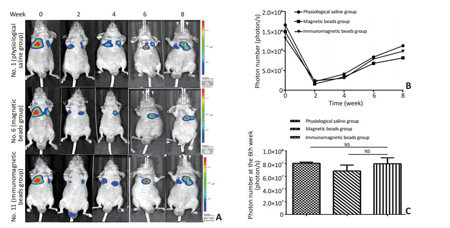
图 1 生物发光成像监测肺部肿瘤的形成和发展
Figure 1. Bioluminescence imaging monitoring the formation and development of lung tumor
A. Each representative mouse of bioluminescent imaging at week 0, 2, 4, 6, 8; B. The bioluminescence imaging signal changes of three groups mice at week 0, 2, 4, 6, 8; C. The comparison of the photon number in three groups mice at the 6th week

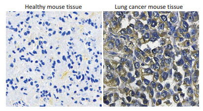
图 2 免疫磁珠悬液对裸鼠肺部肿瘤的早期诊断
Figure 2. Early diagnosis of lung cancer in nude mice after injection of immunomagnetic bead solution

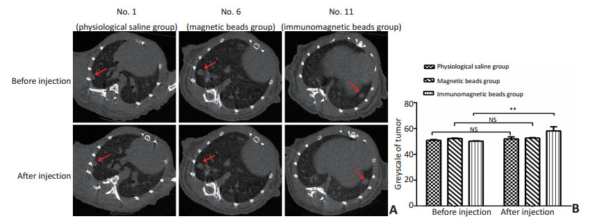
图 3 免疫磁珠溶液注射前后micro-CT扫描的信号变化
A. The largest tumor volume of each representative mouse detected before and after injection by micro-CT scan at the 6th week; B. The comparison of the mouse tumor density before and after injection at the 6th week, **P<0.01
Figure 3. Signal change of micro-CT scan after injection of immunomagnetic bead solution
-
[1] Tsai CH, Li CH, Cheng YW, et al.The inhibition of lung cancer cell migration by AhR-regulated autophagy[J].Scientific Reports, 2017, 7:41927. doi: 10.1038/srep41927 [2] Garg PK, Singh SK, Prakash G, et al.Role of positron emission tomography-computed tomography in non-small cell lung cancer[J].World Journal of Methodology, 2016, 6(1):105-111. doi: 10.5662/wjm.v6.i1.105 [3] van der Aalst CM, Ten HK, de Koning HJ.Lung cancer screening: latest developments and unanswered questions[J].Lancet Respiratory Medicine, 2016, 4(9):749-761. doi: 10.1016/S2213-2600(16)30200-4 [4] Correia LL, Johnson JA, McErlean P, et al.SOX2 drives bronchial dysplasia in a novel organotypic model of early human squamous lungcancer[J].American Journal of Respiratory and Critical Care Medicine, 2017, 195(11):1494-1508. doi: 10.1164/rccm.201510-2084OC [5] Fumagalli C, Bianchi F, Raviele PR, et al.Circulating and tissue biomarkers in early-stage non-small cell lung cancer[J].Ecancermedicalscience, 2017, 195(11):1494-1508. http://www.jtcvsonline.org/article/S0022-5223(01)33231-2/pdf [6] Klupczynska A, Dereziński P, Matysiak J, et al.Determination of 16serum angiogenic factors in stage Ⅰ non-small cell lung cancer usinga bead-based multiplex immunoassay[J].Biomed Pharmacother, 2017, 88:1031-1037. doi: 10.1016/j.biopha.2017.01.141 [7] Ağababaoğlu İ, Önen A, Demir AB, et al.Chaperonin(HSP60) andannexin-2 are candidate biomarkers for non-small cell lung carcinoma[J].Medicine(Baltimore), 2017, 96(6):e5903. https://www.researchgate.net/publication/313893575_Chaperonin_HSP60_and_annexin-2_are_candidate_biomarkers_for_non-small_cell_lung_carcinoma [8] 徐婷, 潘世扬, 王芳, 等.抗人非小细胞肺癌单克隆抗体的制备及鉴定[J].临床检验杂志, 2011, 29(3):216-218. http://cpfd.cnki.com.cn/Article/CPFDTOTAL-ZHYX201105003295.htmXu T, Pan SY, Wang F, et al.Preparation and identification of monoclonal antibody against human non-small cell lung cancer[J].Chinese Journal of Clinical Laboratory Science, 2011, 29(3):216-218. http://cpfd.cnki.com.cn/Article/CPFDTOTAL-ZHYX201105003295.htm [9] 彭蘡, 潘世扬, 王芳, 等.非小细胞肺癌患者血清中SP70的检测及其临床意义[J].中华检验医学杂志, 2012, 35(6):554-558. http://cdmd.cnki.com.cn/Article/CDMD-87112-2009055183.htmPeng Y, Pan SY, Wang F, et al.A preliminary study on serum proteinSP70 as a novel biomarker for the detection of non-small cell lungcancer[J].Chinese Journal of Laboratory Medicine, 2012, 35(6):554-558. http://cdmd.cnki.com.cn/Article/CDMD-87112-2009055183.htm [10] 杨瑞霞, 潘世扬, 王芳, 等.良恶性胸腔积液鉴别中SP70检测的临床意义[J].中华检验医学杂志, 2012, 35(12):1150-1154. doi: 10.3760/cma.j.issn.1009-9158.2012.12.021Yang RX, Pan SY, Wang F, et al.The significance of protein SP70 detection for differentiating benign and malignant pleural effusion[J].Chinese Journal of Laboratory Medicine, 2012, 35(12):1150-1154. doi: 10.3760/cma.j.issn.1009-9158.2012.12.021 [11] Bigbee WL, Gopalakrishnan V, Weissfeld JL, et al.A multiplexed serum biomarker immunoassay panel discriminates clinical lung cancer patients from high-risk individuals found to be cancer-free byCT screening[J].J Thorac Oncol, 2012, 7(4):698-708. doi: 10.1097/JTO.0b013e31824ab6b0 [12] Khiewvan B, Torigian DA, Emamzadehfard S, et al.An update on therole of PET/CT and PET/MRI in ovarian cancer[J].Eur J Nucl MedMol Imaging, 2017, 44(6):1079-1091. doi: 10.1007/s00259-017-3638-z [13] Chen L, Wu X, Ma X, et al.Prognostic value of 18F-FDG PET-CTbased functional parameters in patients with soft tissue sarcoma:A meta-analysis[J].Medicine(Baltimore), 2017, 96(6):e5913. https://www.researchgate.net/publication/313893653_Prognostic_value_of_18F-FDG_PET-CT-based_functional_parameters_in_patients_with_soft_tissue_sarcoma_A_meta-analysis [14] 雍亚兰, 李雪梅, 谭兰英.CT增强扫描造影剂渗漏的预防及护理[J].内蒙古中医药, 2012, 31(15):177-178. doi: 10.3969/j.issn.1006-0979.2012.15.214Yong YL, Li XM, Tan LY.CT enhanced screening for the preventionand care of screening imaging agents[J].Nei Mongol Journal of Traditional Chinese Medicine, 2012, 31(15):177-178. doi: 10.3969/j.issn.1006-0979.2012.15.214 [15] Yin WJ, Yi YH, Guan XF, et al.Preprocedural prediction model forcontrast-induced nephropathy patients[J].J Am Heart Assoc, 2017, 6(2):pii:e004498. [16] Lusic H, Grinstaff M W.X-ray-computed tomography contrast agents[J].Chemical Reviews, 2012, 113(3):1641-1666. [17] 张晓婷, 戴志飞.多功能纳米CT造影剂的研究进展[J].科学通报, 2015, 60:3424-3437. http://www.cnki.com.cn/Article/CJFDTOTAL-KXTB201535004.htmZhang XT, Dai ZF.Advances in multifunctional nano-sized CT contrast agents[J].Chinese Science Bulletin, 2015, 60:3424-3437. http://www.cnki.com.cn/Article/CJFDTOTAL-KXTB201535004.htm [18] Pan S, Wang F, Huang P, et al.The study on newly developed McAbNJ001 specific to non-small cell lung cancer and its biological characteristics[J].Plos One, 2012, 7(3):e33009. doi: 10.1371/journal.pone.0033009 [19] Allen TM, Cullis PR.Drug delivery systems: entering the mainstream[J].Science, 2004, 303(5665):1818-2182. doi: 10.1126/science.1095833 [20] 李大千, 吴蕾, 荆俊鹏, 等.生物发光成像对裸鼠肺部转移瘤的早期检测研究[J].南京医科大学学报(自然科学版), 2015, 35(11):1522-1527. http://www.cnki.com.cn/Article/CJFDTOTAL-NJYK201511005.htmLi DQ, Wu L, Jing JP, et al.Application of bioluminescence imagingon early intrapulmonary micrometastases tumors in mouse model[J].Acta Universitatis Medicinalis Nanjing, 2015, 35(11):1522-1527. http://www.cnki.com.cn/Article/CJFDTOTAL-NJYK201511005.htm -

 下载:
下载:

 点击查看大图
计量
- 文章访问数: 53
- HTML全文浏览量: 11
- PDF下载量: 3
- 被引次数: 0
