
Comparison of rectal cancer tumor volume parameters measured by MRI sequences and CT with those by pathological specimen
-
摘要:
目的 比较直肠癌术后病理标本与术前MRI(T1WI、T2WI、DWI)和/或CT显示的肿瘤范围的差异。方法:选取2016年3月至2016年5月间于湖南省肿瘤医院拟行全直肠系膜切除术(TME)的直肠癌患者22例,术前行盆腔MRI(T1WI、T2WI、DWI)和/或增强CT检查。分别测量肿瘤沿肠管纵轴长度、垂直肠管横轴位肿瘤最宽径和横轴位肿瘤实际面积,并与术后病理标本测量对应参数比较,评估各影像测量的精确性。 结果 病理长度(Lpath-L)为(4.06±1.14)cm,LT1-L、LT2-L、LDWI-L、LCT-L分别为(3.91±1.51)、(4.62±1.41)、(3.39±1.05)、(3.94±1.23)cm,与Lpath-L的相关系数分别为0.688、0.635、0.688、0.720(P < 0.05);T2WI测量结果存在平均6 mm高估,T1WI、DWI、CT测量结果存在1~6 mm不同程度的低估。病理横截面肿瘤最宽径(Lpath-W)为(2.56±0.94)cm,LT1-W、LT2-W、LDWI-W、LCT-W分别为(3.62±0.99)、(3.66±0.76)、(3.23±0.58)、(3.64±1.04)cm,测量结果存在平均5.1~11.1 mm的高估。肿瘤病理实际面积(Apath)为(4.30±2.83)cm2,AT1、AT2、ADWI、ACT分别为(8.98±3.90)、(8.99±3.43)、(8.41±3.09)、(9.63±4.40)cm2,各影像测量实际面积存在约2倍程度高估。结论:在病变长度方面,各影像存在-6~6 mm差异;最大横截面方面均存在不同程度高估。因此在直肠癌放疗GTV勾画时,断面侧方应适当保守内收,而上下端也不应过多延伸。 Abstract:Objective This study aimed to compare rectal cancer tumor volume parameters measured by MRI sequences (T1WI, T2WI, and DWI) and/or CT with those by pathological specimen. Methods Twenty-two patients with rectal cancer were prospectively enrolled. MRI sequences including T1WI, T2WI, and DWI, and/or CT of the pelvis were performed before operation. Volume parameters, such as tumor length along the rectal axis, maximum tumor width perpendicular to rectal axis, and tumor actual area in that perpendicular plane, were measured on T1WI, T2WI, DWI, and CT, respectively, for each patient. The respective pathological parameters were further measured in surgical specimen after total mesorectal excision. The two kinds of parameter values measured in imaging and pathology were statistically compared and accuracy appraisal was performed. Results The mean Lpath-L was 4.06±.14 cm. The mean LT1-L, LT2-L, LDWI-L, and LCT-L were 3.91±1.51, 4.62±.41, 3.39±.05, and 3.94±.23 cm, respectively. Correlation coefficients were 0.688, 0.635, 0.688, and 0.720 (P < 0.05). An average 6 mm overestimation was found in T2WI, and 1 to 6 mm underestimation in T1WI, DWI, and CT in length values compared with those measured in surgical specimen. The mean Lpath-W was 2.56 ±.94 cm. The mean LT1-W, LT2-W, LDWI-W, and LCT-W were 3.62±.99, 3.66±.76, 3.23±.58, and 3.64±.04 cm, respectively. The magnitude of mean overestimation ranged from 5.1 to 11.1 mm. The Apath was 4.30 ±.83 cm2. AT1, AT2, ADWI, and ACT were 8.98±.90, 8.99±.43, 8.41±.09, and 9.63±.40 cm2, respectively, which double overestimated the tumor area in the perpendicular rectal plane. Conclusion The difference in longitudinal length between MRI sequences/CT and pathological specimen was in the range of -6 mm to 6 mm. The mean maximum tumor width and areas in the maximum tumor perpendicular plane were overestimated. This study indicated that gross tumor volume delineation based on CT or MRI for rectal cancer irradiation should be conservative in the axial images of rectum, and meticulous consideration is required along the rectum. -

图 1 直肠癌患者不同影像大体肿瘤体积(GTV)勾画
Figure 1. Example of the gross tumor volume (GTV) delineation in a rectal cancer patient
A. Sagittal T2WI; B. CT; C. Axial T2WI; D. DWI; E. LAVA-T1WI. Of the same patient, the tumor contour presented in red

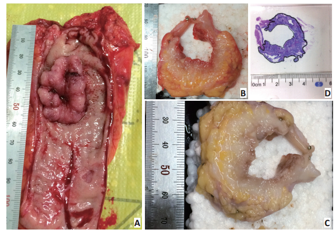
图 2 直肠癌病理标本的处理和肿瘤体积参数测量
Figure 2. Processing of pathological specimen and measurement of tumor volume parameters for rectal cancer
A. Measurement of the gross tumor length immediately after total mesorectum excision; B. Slicing across the widest tumor region with 5 mm thickness perpendicular to the rectum axis, recovering the specimen slice to its original form on the hollow board and fixing onto it with pins; C. Formalin specimen fixation for 24 hours; D. The slice was made into slides and stained with hematoxylin and eosin, and the tumor-containing boundary was macroscopically outlined with microscope

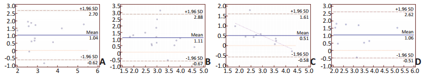
图 3 不同影像测量与病理测量最大横截面肿瘤最宽径的Bland-Altman一致性分析
Figure 3. Bland-Altman plots showing the difference against mean maximum tumor width perpendicular to rectal axis measured on three imaging modalities and pathological parameters
A. T1WI; B. T2WI; C. DWI; D. CT. Dashed lines represent the 95% confidence intervals around the mean of difference. The solid lines represent the mean of difference
表 1 22例直肠癌患者不同影像测量的大体肿瘤体积长度与术后标本病变长度的Pearson相关分析
Table 1. Correlation between rectal tumor length measured by MRI sequences (T1WI, T2WI, DWI), CT, and by pathological specimen

-
[1] Araujo RO, Valadão M, Borges D, et al. Nonoperative management of rectal cancer after chemoradiation opposed to resection after complete clinical response. A comparative study[J]. Eur J Surg Oncol, 2015, 41(11):1456-1463. doi: 10.1016/j.ejso.2015.08.156 [2] Lee SY, Kim CH, Kim YJ, et al. Oncologic outcomes according to the treatment strategy in radiologic complete responders after neoadjuvant chemoradiation for rectal cancer[J]. Oncology, 2015, 89(6): 311-318. doi: 10.1159/000439279 [3] Li J, Liu H, Yin J, et al. Wait-and-see or radical surgery for rectal cancer patients with a clinical complete response after neoadjuvant chemoradiotherapy: a cohort study[J]. Oncotarget, 2015, 6(39): 42354-42361. doi: 10.18632/oncotarget.v6i39 [4] Appelt AL, Pløen J, Harling H, et al. High-dose chemoradiotherapy and watchful waiting for distal rectal cancer: a prospective observational study[J]. Lancet Oncol, 2015, 16(8):919-927. doi: 10.1016/S1470-2045(15)00120-5 [5] Daisne JF, Duprez T, Weynand B, et al. Tumor volume in pharyngolaryngeal squamous cell carcinoma: a comparison at CT, MR imaging and FDG PET and validation with surgical specimen[J]. Radiology, 2004, 233(1):93-100. doi: 10.1148/radiol.2331030660 [6] Han DL, Yu JM, Yu YH, et al. Comparison of 18F-Fluorothymidine and 18F-Fluorodeoxyguocose PET/CT in delineating gross tumor volume by optimal threshold in patients with squamous cell carcinoma of thoracic esophagus[J]. Int J Radiat Oncol Biol Phys, 2010, 76 (4):1235-1241. doi: 10.1016/j.ijrobp.2009.07.1681 [7] Yu JM, Li XK, Xing LG, et al. Comparison of tumor volumes as determined by pathologic examination and FDG-PET/CT images of nonsmall-cell lung cancer: a pilot study[J]. Int J Radiation Oncology Biol Phys, 2009, 75(5):1468-1474. doi: 10.1016/j.ijrobp.2009.01.019 [8] Stroom J, Blaauwgeers H, van Baardwijk A, et al. Feasibility of pathology-correlated lung imaging for accurate target definition of lung tumors[J]. Int J Radiation Oncology Biol Phys, 2007, 69(1):267-275. doi: 10.1016/j.ijrobp.2007.04.065 [9] Buijsen J, van den Bogaard J, Janssen MH. et al. FDG-PET provides the best correlation with the tumor specimen compared to MRI and CT in rectal cancer.[J] Radiother and Oncol, 2011, 98(2):270-276. doi: 10.1016/j.radonc.2010.11.018 [10] Liao CY, Chen SW, Wu YC, et al.Correlations between 18F-FDG PET/ CT parameters and pathological findings in patients with rectal cancer[J]. Clin Nucl Med, 2014, 39(1):40-45. doi: 10.1097/RLU.0b013e318292f0f6 [11] Feng Q, Yan YQ, Zhu J, et al. T staging of rectal cancer: accuracy of diffusion-weighted imaging compared with T2-weighted imaging on 3.0 tesla MRI[J]. J Dig Dis, 2014, 15(4):188-194. doi: 10.1111/cdd.2014.15.issue-4 [12] van der Paardt MP, Zagers MB, Beets-Tan RG, et al. Patients who undergo preoperative chemoradiotherapy for locally advanced rectal cancer restaged by using diagnostic MR imaging: a systematic review and meta-analysis[J]. Radiology, 2013, 269(1):101-112. doi: 10.1148/radiol.13122833 [13] Burbach JP, Kleijnen JP, Reerink O, et al. Inter-observer agreement of MRI-based tumor delineation for preoperative radiotherapy boost in locally advanced rectal cancer[J]. Radiother Oncol, 2016, 118(2):399-407. doi: 10.1016/j.radonc.2015.10.030 [14] Jonmarker S, Valdman A, Lindberg A, et al. Tissue shrinkage after fixation with formalin injection of prostatectomy specimens[J]. Virchows Arch, 2006, 449(3):297-301. doi: 10.1007/s00428-006-0259-5 [15] Caldas-Magalhaes J, Kasperts N, Kooij N, et al. Validation of imaging with pathology in laryngeal cancer: accuracy of the registration methodology[J]. Int J Radiat Oncol Biol Phys, 2012, 82(2):289-298. doi: 10.1016/j.ijrobp.2011.05.004 [16] Brændengen M, Hansson K, Radu C, et al. Delineation of gross tumor volume (GTV) for radiation treatment planning of locally advanced rectal cancer using information from MRI or FDG-PET/CT: a prospective study[J]. Int J Radiation Oncology Biol Phys, 2011, 81 (4):439-445. doi: 10.1016/j.ijrobp.2011.03.031 [17] 侯栋梁, 时高峰, 高献书等.磁共振弥散加权成像在食管癌大体肿瘤靶区勾画中的应用价值[J].中华放射肿瘤学杂志, 2012, 21(4), 343-347. http://cdmd.cnki.com.cn/Article/CDMD-10089-1014248047.htmHou DL, Shi GF, Gao XS, et al. The application value of diffusionweighted magnetic resonance imaging in gross tumor volumedelineation ofesophageal squamous cell carcinoma[J]. ChinJ Radiat Oncol, 2012, 21(4):343-347. http://cdmd.cnki.com.cn/Article/CDMD-10089-1014248047.htm -

 下载:
下载:
 点击查看大图

计量
- 文章访问数: 114
- HTML全文浏览量: 77
- PDF下载量: 3
- 被引次数: 0
