
-
摘要:
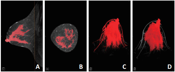
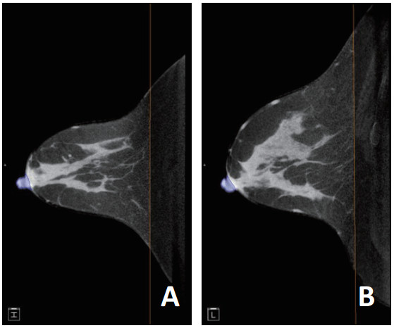
目的 探讨基于锥光束乳腺CT(cone beam breast CT,CBBCT)的阈值分割法测量乳腺密度的准确性,及其对乳腺腺体分类和乳腺癌筛查的意义。 方法 回顾性分析2012年5月至2013年9月于天津医科大学肿瘤医院行乳腺X线检查(mammography,MG)及CBBCT检查的195例患者的影像学资料,其中64例患者的64侧乳腺符合入组条件。依据BI-RADS中乳腺构成的分类标准对其进行分类并得到多数报告;基于其CBBCT图像进行阈值分割法测量乳腺密度,并得到手动修正后乳腺密度。1个月后重复上述步骤。采用组内相关系数(intraclass correlation coefficient,ICC)比较观察者内、观察者间、阈值分割法测量与手动修正、非致密类及致密类乳腺测量结果之间的一致性。 结果 阈值分割法测量乳腺密度的观察者内和观察者间ICC值分别为0.9624(95% CI:0.9388~0.9770)和0.9666(95%CI:0.9500~0.9785);手动修正测量观察者内和观察者间ICC值分别为0.9750(95%CI:0.9592~ 0.9847)和0.9775(95%CI:0.9661~0.9855);阈值分割法与手动修正测量之间ICC值为0.9962(95%CI:0.9983~0.9977);非致密类和致密类乳腺阈值分割法与手动修正之间ICC值分别为0.9497(95%CI:0.7072~0.9914)和0.9983(95%CI:0.9971~0.9990)。 结论 基于CBBCT图像的阈值分割法是一种较为稳定且准确的计算机辅助测量乳腺密度的方法,未来有望应用于大规模乳腺癌筛查,并为乳腺癌风险的预测提供更多信息。 Abstract:Objective To investigate the accuracy of a threshold-based segmentation method based on cone beam breast CT (CBBCT) images in breast density measurement, and its value for breast-type classification and breast cancer screening. Methods A retrospective analysis of 195 patients who had undergone CBBCT examination at Tianjin Medical University Cancer Institute and Hospital between May 2012 and August 2014 was performed. A total of 64 breasts were analyzed. On the basis of the classification criteria for breast density in BI-RADS, they were classified into four types and the majority report was reported. Breast density was measured by the threshold-based segmentation method based on CBBCT images and corrected manually to obtain the corrected breast density. A month later, the procedure was repeated. Intra-class correlation coefficients (ICCs) were used to compare the intra-observer and interobserver consistencies of threshold-based segmentation and manually corrected breast density measurement results for non-dense and dense breasts. Results For threshold-based segmentation measurements the intra-observer and inter-observer ICC values were 0.0.9624 (95% CI: 0.9388~0.9770) and 0.9666 (95% CI: 0.9500~0.9785). For manually corrected measurements, the intra-observer and inter-observer ICC values were 0.9750 (95% CI: 0.9592~0.9847) and 0.9775 (95% CI: 0.9661~0.9855). The ICC between the threshold-based segmentation method and manual correction was 0.9962 (95% CI: 0.9983~0.9977). The ICC values of thresholdbased and manually corrected measurement in non-dense and dense breasts were 0.9497 (95% CI:0.7072-0.9914) and 0.9983 (95% CI: 0.9971-0.9990), respectively. Conclusions The threshold-based segmentation method based on CBBCT is a reliable and accurate computer-aided method of measuring breast density. It is expected to be applied in large-scale screening of breast cancer and to provide more information for predicting the risk of breast cancer -
Key words:
- breast density /
- cone beam breast CT /
- breast cancer screening
-
表 1 基于CBBCT图像进行阈值分割法及手动修正测量乳腺密度结果

表 2 阈值分割法及手动修正测量乳腺密度的观察者内及观察者间ICC值

-
[1] Wolfe JN. Breast patterns as an index of risk for developing breast cancer[J]. AJR Am J Roentgenol, 1976, 126(6):1130-1137. doi: 10.2214/ajr.126.6.1130 [2] Baglietto L, Krishnan K, Stone J, et al. Associations of mammographic dense and nondense areas and body mass index with risk of breast cancer[J]. Am J Epidemiol, 2014, 179(4):475-483. doi: 10.1093/aje/kwt260 [3] Peres J. Understanding breast density and breast cancer risk[J]. J Natl Cancer Inst, 2012, 104(18):1345-1346. doi: 10.1093/jnci/djs403 [4] Ahmadinejad N, Movahedinia S, Movahedinia S, et al. Association of mammographic density with pathologic findings[J]. Iran Red Crescent Med J, 2013, 15(12):e16698. http://europepmc.org/abstract/med/24693404 [5] 李海洁, 尹璐, 叶兆祥, 等.锥光束乳腺CT及乳腺X线摄影对乳腺组织覆盖范围的比较[J].中华放射学杂志, 2015, 49(7):488-490. doi: 10.3760/cma.j.issn.1005-1201.2015.07.003 [6] 美国放射学院.乳腺影像报告与数据系统图谱(2013版)[M].北京:北京大学医学出版社, 2016, 116-123. [7] Irshad A, Leddy R, Ackerman S, et al. Effects of changes in BI-RADS density assessment guidelines (fourth versus fifth edition) on breast density assessment: intra-and interreader agreements and density distribution[J]. AJR Am J Roentgenol, 2016, 207(6):1366-1371. doi: 10.2214/AJR.16.16561 [8] Ciatto S, Bernardi D, Calabrese M, et al. A first evaluation of breast radiological density assessment by QUANTRA software as compared to visual classification[J]. Breast, 2012, 21(4):503-506. doi: 10.1016/j.breast.2012.01.005 [9] Redondo A, Comas M, Macià F, et al. Inter-and intraradiologist variability in the BI-RADS assessment and breast density categories for screening mammograms[J]. Br J Radiol, 2012, 85(1019):1465-1470. doi: 10.1259/bjr/21256379 [10] Bernardi D, Pellegrini M, Di Michele S, et al. Interobserver agreement in breast radiological density attribution according to BIRADS quantitative classification[J]. Radiol Med, 2012, 117(4):519-528. doi: 10.1007/s11547-011-0777-3 [11] Dehkordy SF, Carlos RC. Dense breast legislation in the United States: state of the states[J]. J Am Coll Radiol, 2016, 13(11S):R53-R57. http://cn.bing.com/academic/profile?id=13457f06a8897115a13772ba08676da6&encoded=0&v=paper_preview&mkt=zh-cn [12] Brandt KR, Scott CG, Ma L, et al. Comparison of clinical and automated breast density measurements: implications for risk prediction and supplemental screening[J]. Radiology, 2016, 279(3):710-719. doi: 10.1148/radiol.2015151261 [13] 李二妮, 周纯武, 李静, 等.乳腺密度的影响因素及其在乳腺疾病中的诊断价值[J].实用放射学杂志, 2013, 29(10):1570-1572. doi: 10.3969/j.issn.1002-1671.2013.10.005 -

 下载:
下载:

 点击查看大图

图(4) / 表(2)
计量
- 文章访问数: 68
- HTML全文浏览量: 4
- PDF下载量: 1
- 被引次数: 0
