
Application of digital angle instrument in computed tomography-guided percutaneous lung biopsy
-
摘要:
目的 CT引导经皮肺穿刺活检对于肺小结节以及临近大血管的病灶具有难度。本研究旨在探讨数显角度仪在CT引导经皮肺穿刺活检中的应用价值。 方法 回顾性分析2018年1月至2018年9月南华大学附属第一医院经皮肺穿刺活检的35例患者。将患者分为A、B、C3组,A组与B组为病灶直径≤30 mm的肺结节,其中A组接受数显角度仪协助下的CT引导经皮肺穿刺活检,B组则在CT引导下徒手操作经皮肺穿刺活检。C组为病灶直径 > 30 mm肺部肿块,在CT引导下徒手操作经皮肺穿刺活检。然后比较3组之间穿刺前的肿块大小、进针次数、进针距离及术后血气胸并发症的差异。 结果 A组肺结节最大直径为(18.4±2.1)mm,显著小于B组(28.3±2.0)mm及C组(43.3±3.6)mm(P=0.003与P=0.003)。A组部分患者合并严重慢性阻塞性肺疾病(chronic obstructive pulmonary diseases,COPD)及病灶邻近大血管,且该组穿刺点至胸壁外侧距离也明显高于B组(P=0.039)。但A组一次性穿刺成功概率为100%,明显高于B、C组,同时该组术后并发症亦明显少于另外两组。 结论 经数显角度仪协助的CT引导下经皮肺穿刺活检术为一项安全、简便、准确的诊断方法,特别在肺小结节病变的患者中具有较好的应用价值。 Abstract:Objective Computed tomography (CT)-guided percutaneous lung biopsy is difficult for small nodules and lesions that are adjacent to large blood vessels. This study investigated the validity of CT-guided percutaneous lung biopsy in the diagnosis of pulmonary nodules with a digital angle instrument. Methods This study was a retrospective analysis of 35 patients with lung mass ≤60 mm, who underwent CT- guided percutaneous lung biopsy from January 2018 to September 2018. Patients were assigned in to three groups. Group A and B were patients with pulmonary nodules ≤30 mm. Biopsy of group A was performed with the help of a digital angle instrument, and group B didn' t use digital angle instrument. Group C had lung mass of > 30 mm, and the biopsy was performed without using the instrument. The size of the mass, frequency of punctures, distance of the puncture, and complication of pneumothorax after puncture were compared among the three groups. Results The maximum diameter of pulmonary nodules in group A (18.4 ± 2.1) mm was significantly lower than that in groups B (28.3 ± 2.0) mm and C (43.2 ± 3.6) mm, and their P value were 0.0034 and 0.0028, respectively. Some patients in group A were at risk because of severe chronic obstructive pulmonary disease and proximity of lesions to large blood vessels. The puncture distance in group A was also significantly more than groups B (P < 0.039). However, the probability of puncture success in group A was 100%, which was significantly higher than groups B and C. The postoperative complications in group A were also significantly fewer than in other two groups. Conclusions CT-guided percutaneous lung biopsy with a digital angle instrument is a safe, simple, and accurate diagnostic method, especially in patients with pulmonary nodular lesions. -
Key words:
- lung nodules /
- lung cancer /
- lung biopsy /
- digital angle instrument
-

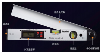
图 2 数显角度仪协助CT引导下经皮肺穿刺活检术的具体步骤
▶A:裁剪不锈钢网;B:CT初步定位;C:确定体表进针位置、角度及深度;D:棉签在患者体表印迹,方便消毒后进针;E:数显角度仪协助穿刺进针;F:活检针进入肿瘤边缘;G:活检;H:活检完毕再次行CT扫描,观察有无气胸和出血发生;I:病理学检查(H & E×100);C、F:箭头提示肺内病灶,三角形为穿刺活检针

图 3 3组患者行穿刺活检的肿块大小、进针距离及穿刺次数比较
A:肿块最大直径的比较;B:病灶穿刺点到外侧胸壁的距离比较;C:病灶穿刺点到内侧胸壁的距离比较;D:穿刺活检进针次数比较

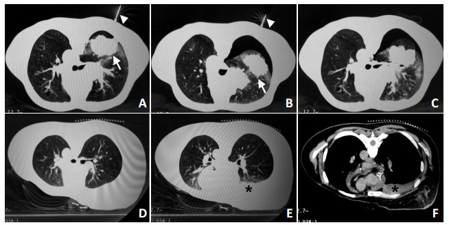
图 4 2例患者接受数显角度仪协助CT引导下行经皮肺穿刺活检
A:1例患者穿刺前CT图像;B:图A患者行经皮肺穿刺活检,显示穿刺针达到肿瘤边缘;C:另1例患者肺结节仅5 mm,在穿刺过程中再次行CT检查,通过其指导后续进针角度及深度;D:图C患者行经皮肺穿刺活检的穿刺针到达肿瘤边缘

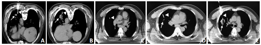
图 5 2例患者CT引导下徒手操作经皮肺穿刺活检术的多次进针
A:1例患者首次穿刺活检,穿刺针未在肿块内;B:图A患者行第2次穿刺活检,显示穿刺针到达肿瘤边缘;C:另1例患者首次穿刺活检,穿刺针未在肿块内;D:图C患者第2次穿刺活检,穿刺针仍未在肿块内;E:图C患者第3次穿刺活检,显示穿刺针到达肿瘤边缘

图 6 CT引导下徒手操作经皮肺穿刺活检术术后并发症
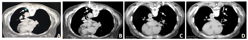
A:1例患者行穿刺活检,穿刺针刚进入肿块内,未见气胸;B:图A患者行穿刺中,开始出现气胸;C:图A患者行穿刺结束,再次行CT,提示左肺少到中量气胸;D:另1例患者穿刺前,未见血胸;E:图D患者穿刺后,出现血胸肺窗图;F:患者行穿刺后,出现血胸纵膈窗图;箭头提示肺内病灶,三角提示穿刺活检针;*:提示穿刺之后血胸形成
表 1 35例患者接受经皮肺穿刺活检术后的病理结果

-
[1] Chen W, Zheng R, Baade PD, et al. Cancer statistics in China, 2015 [J]. CA Cancer J Clin, 2016, 66(2):115-132. doi: 10.3322/caac.21338 [2] International Early Lung Cancer Action Program Investigators. Survival of patients with stageⅠ lung cancer detected on CT screening [J]. N Engl J Med, 2006, 355(17):1763-1771. doi: 10.1056/NEJMoa060476 [3] Lu C, Onn A, Vaporciyan AA. Holland-frei cancer medicine (8th)[M]. People's Medical Publishing House, USA, 2010:999-1043. [4] Ost D, Fein AM, Feinsilver SH. CLinical practice. The solitary pulmonary nodule[J]. N Engl J Med, 2003, 348(25):2535-2542. doi: 10.1056/NEJMcp012290 [5] Jude CM, Nayak NB, Patel MK, et al. Pulmonary coccidioidomycosis: pictorial review of chest radiographic and CT findings[J]. Radiographics, 2014, 34(4):912-925. doi: 10.1148/rg.344130134 [6] Mukhopadhyay S. Utility of small biopsies for diagnosis of lung nodules: doing more with less[J]. Modern Pathology, 2012, 25(S1):543-557. http://cn.bing.com/academic/profile?id=691a5debb88acb3595678ca99b677411&encoded=0&v=paper_preview&mkt=zh-cn [7] Heerink WJ, de Bock GH, de Jonge GJ, et al. Complication rates of CT-guided transthoracic lung biopsy: meta-analysis[J]. European radiology, 2017, 27(1):138-148. doi: 10.1007/s00330-016-4357-8 [8] Winer-Muram HT. The solitary pulmonary nodule[J]. Radiology, 2006, 239(1):34-49. doi: 10.1148/radiol.2391050343 [9] Yasaka K, Katsura M, Hanaoka S, et al. High-resolution CT with new model-based iterative reconstruction with resolution preference algorithm in evaluations of lung nodules: Comparison with conventional model-based iterative reconstruction and adaptive statistical iterative reconstruction[J]. Eur J Radiol, 2016, 85(3):599-606. doi: 10.1016/j.ejrad.2016.01.001 [10] Liu Y, Wang H, Li Q, et al. Radiologic features of small pulmonary nodules and lung cancer risk in the national lung screening trial: a nested case-control study[J]. Radiology, 2017, 286(1):298-306. http://cn.bing.com/academic/profile?id=74256ef6d819291a9e3cb6dc45f87d2d&encoded=0&v=paper_preview&mkt=zh-cn [11] Yang W, Sun W, Li Q, et al. Diagnostic accuracy of CT-guided transthoracic needle biopsy for solitary pulmonary nodules[J]. PLoS One, 2015, 10(6):e0131373. doi: 10.1371/journal.pone.0131373 [12] Li GC, Fu YF, Cao W, et al. Computed tomography-guided percutaneous cutting needle biopsy for small (≤20 mm) lung nodules[J]. Medicine, 2017, 96(46):e8703. doi: 10.1097/MD.0000000000008703 [13] Sachdeva M, Ronaghi R, Mills PK, et al. Complications and yield of computed tomography-guided transthoracic core needle biopsy of lung nodules at a high-volume academic center in an endemic coccidioidomycosis area[J]. Lung, 2016, 194(3):379-385. doi: 10.1007/s00408-016-9866-3 [14] Hwang HS, Chung MJ, Lee JW, et al. C-arm cone-beam CT-guided percutaneous transthoracic lung biopsy: usefulness in evaluation of small pulmonary nodules[J]. Am J Roentgenol, 2010, 195(6):W400- W407. doi: 10.2214/AJR.09.3963 [15] 肖天林, 徐细明, 戈伟.三维立体定向仪在肺内小结节经皮活检中的应用[J].武汉大学学报(医学版), 2015, 36(6):922-925. http://d.old.wanfangdata.com.cn/Periodical/hubeiykdxxb201506019 -

 下载:
下载:


 点击查看大图
计量
- 文章访问数: 133
- HTML全文浏览量: 8
- PDF下载量: 6
- 被引次数: 0
