
The clinical value of endobronchial ultrasound elastography in the diagnosis of mediastinal and hilar lymph nodes
-
摘要:
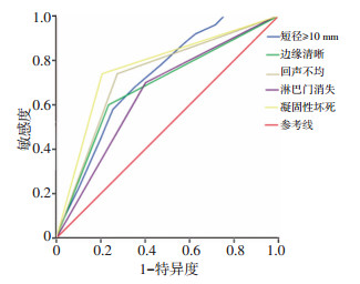
目的 用4种支气管内超声实时弹性成像(endobronchial ultrasound real-time elastography,EBUS-RTE)评定方法对比分析纵隔及肺门淋巴结的弹性成像图,探讨其在纵隔及肺门淋巴结良恶性鉴别诊断中的临床价值。 方法 选择2016年1月至2018年12月在兰州大学第二医院接受支气管内超声引导下的经支气管针吸活检(endobronchial ultrasound-guided transbronchial needle aspiration,EBUS-TBNA)检查的患者,同时行EBUS-RTE操作。分别使用弹性图像类型、弹性评分、应变率比值(strain ratio,SR)及蓝色区域面积比例法(blue color proportion,BCP)分析淋巴结的弹性成像图,最后以EBUS-TBNA的病理结果、微生物检测结果确定病变淋巴结的性质。为评价4种方法的诊断价值,建立受试者工作特征曲线(receiver operating characteristic,ROC),计算出曲线下面积(area under curve,AUC)及最佳诊断临界值,同时比较4种方法的敏感度、特异度、阳性预测值、阴性预测值及准确率。 结果 共入组80例患者,152枚纵隔/肺门淋巴结,其中102枚恶性淋巴结,50枚良性淋巴结。弹性图像类型、弹性评分、应变率比值及BCP 4种评定方法在淋巴结良恶性鉴别诊断中差异均有统计学意义(均P < 0.01)。统计结果显示,BCP诊断良恶性淋巴结的准确率最高为88.3%,诊断良恶性淋巴结的敏感度、特异度、阳性预测值及阴性预测值分别为90.0%、94.1%、85.4%和88.6%。BCP的最佳界定值为85.7%,BCP≥85.7%的65枚淋巴结均为恶性淋巴结。 结论 弹性图像类型、弹性评分、应变率比值及BCP 4种超声弹性成像评定方法对纵隔及肺门淋巴结良恶性的鉴别诊断具有较高的临床应用价值,而BCP的诊断价值最高。 Abstract:Objective Four ultrasound elastography parameters were used to compare the color distribution of mediastinal and hilar lymph nodes (LNs) in endobronchial ultrasound real-time elastography (EBUS-RTE), and the clinical value in differentiating benign from malignant mediastinal and hilar lymph nodes were explored. Methods We selected patients who received EBUS-TBNA in the Second Hospital of Lanzhou University from January 2016 to December 2018 and simultaneously conducted EBUS-RTE. Elastography of lymph nodes were analyzed by elastography image type, elasticity score, strain ratio (SR), and blue color proportion (BCP), respectively. A receiver operating characteristic (ROC) curve was constructed to evaluate the diagnostic value of the four ultrasound elastography parameters. The area under the curve (AUC) and the critical value of the best diagnosis were calculated, and the sensitivity, specificity, positive predictive value, negative predictive value, and accuracy of the four parameters were compared. Results A total of 80 patients were enrolled, including 152 lymph nodes with 102 malignant lymph nodes and 50 benign ones. Elastography image type, elasticity score, SR, and BCP had statistical significance in the differential diagnosis of benign and malignant lymph nodes (P < 0.01). The accuracy of BCP in the diagnosis of benign and malignant lymph nodes was the highest (88.3%), and the sensitivity, specificity, positive predictive value, and negative predictive value were 90.0%, 94.1%, 85.4%, and 88.6%, respectively. The optimal definition value of BCP was 85.7%; all the 65 LNs with a BCP ≥85.7% were diagnosed as malignant. Conclusions Elastography image type, elasticity score, SR, and BCP of ultrasound elastography are important in the differential diagnosis of mediastinal and hilar lymph nodes, and BCP showed the highest diagnostic valuation. -
表 1 EBUS-RTE各参数在良恶性淋巴结中的特征分析

表 2 EUS及EBUS-RTE参数的诊断价值比较 (%)

-
[1] Dietrich CF, Annema JT, Clementsen P, et al. Ultrasound techniques in the evaluation of the mediastinum, partⅠ: Endoscopic ultrasound (EUS), endobronchial ultrasound (EBUS) and transcutaneous mediastinal ultrasound (TMUS), introduction into ultrasound techniques[J]. J Thoracic Dis, 2015, 7(9):E311-E325. [2] Jenssen C, Annema JT, Clementsen P, et al. Ultrasound techniques in the evaluation of the mediastinum, part 2: mediastinal lymph node anatomy and diagnostic reach of ultrasound techniques, clinical work up of neoplastic and inflammatory mediastinal lymphadenopathy using ultrasound techniques and how to learn mediastinal endosonography[J]. J Thoracic Dis, 2015, 7(10):E439-E458. http://cn.bing.com/academic/profile?id=64c236e54c6235c70b54f557b1898451&encoded=0&v=paper_preview&mkt=zh-cn [3] Ettinger DS, Wood DE, Akerley W, et al. Non-small cell lung cancer, Version 62015[J]. J Natl Compr Canc Netw, 2015, 13 (5):515-524. doi: 10.6004/jnccn.2015.0071 [4] Krouskop TA, Wheeler TM, Kallel F, et al. Elastic moduli of breast and prostate tissues under compression[J]. Ultrasonic imaging, 1998, 20(4): 260-274. doi: 10.1177/016173469802000403 [5] Trosini-Désert V, Jeny F, Taillade L, et al. Bronchial endoscopic ultrasound elastography: preliminary feasibility data[J]. Eur Respir J, 2013, 41(2):477-479. doi: 10.1183/09031936.00124812 [6] Izumo T, Sasada S, Chavez C, et al. Endobronchial ultrasound elastography in the diagnosis of mediastinal and hilar lymph nodes[J]. Jpn J Clin Oncol, 2014, 44(10):956-962. doi: 10.1093/jjco/hyu105 [7] He HY, Huang M, Zhu J, et al. Endobronchial ultrasound elastography for diagnosing mediastinal and hilar lymph nodes[J]. Chin Med J, 2015, 128 (20):2720-2725. doi: 10.4103/0366-6999.167296 [8] Lin CK, Chang LY, Yu KL, et al. Differentiating metastatic lymph nodes in lung cancer patients based on endobronchial ultrasonography features[J]. Med Ultrason, 2018, 20(2):154-158. doi: 10.11152/mu-1282 [9] Rozman A, Malovrh MM, Adamic K, et al. Endobronchial ultrasound elastography strain ratio for mediastinal lymph node diagnosis[J]. Radiol Oncol, 2015, 49(4):334-340. doi: 10.1515/raon-2015-0020 [10] Ma H, An Z, Xia P, et al. Semi-quantitative analysis of EBUS elastography as a feasible approach in diagnosing mediastinal and hilar lymph nodes of lung cancer patients[J]. Scientific reports, 2018, 8(1):3571-3578. doi: 10.1038/s41598-018-22006-4 [11] 杨会珍, 滕家俊, 钟润波, 等.气管内超声引导下经支气管针吸活检对肺内病变的诊断价值[J].中华结核和呼吸杂志, 2013, 36(1):17-21. doi: 10.3760/cma.j.issn.1001-0939.2013.01.009 [12] Goldstraw P, Chansky K, Crowley J, et al. The IASLC lung cancer staging project: proposals for revision of the TNM stage groupings in the forthcoming (eighth) edition of the TNM classification for lung cancer [J]. J Thoracic oncol, 2016, 11(1):39-51. http://cn.bing.com/academic/profile?id=f98b52ba6957ba6a7017df2c1245539d&encoded=0&v=paper_preview&mkt=zh-cn [13] Nakajima T, Inage T, Sata Y, et al. Elastography for predicting and localizing nodal metastases during endobronchial ultrasound[J]. Respiration, 2015, 90(6):499-506. doi: 10.1159/000441798 [14] Furukawa MK, Kubota A, Hanamura H, et al. Clinical application of realtime tissue elastography to head and neck cancer- evaluation of cervical lymph node metastasis with real-time tissue elastography[J]. Nippon Jibiinkoka Gakkai Kaiho, 2007, 110(7):503-505. doi: 10.3950/jibiinkoka.110.503 [15] Itoh A, Ueno E, Tohno E, et al. Breast disease: clinical application of US elastography for diagnosis[J]. Radiology, 2006, 239(2):341-350. doi: 10.1148/radiol.2391041676 [16] Korrungruang P. Diagnostic value of endobronchial ultrasound elastography for the differentiation of benign and malignant intrathoracic lymph nodes[J]. Respiro logy (Carlton, Vic), 2017, 22(5):972-977. doi: 10.1111/resp.12979 -

 下载:
下载:

 点击查看大图
图(2) / 表(2)
计量
- 文章访问数: 57
- HTML全文浏览量: 27
- PDF下载量: 4
- 被引次数: 0
