
Correlation between MRI features and tumor risk grade in gastrointestinal stromal tumors
-
摘要:
目的 分析胃肠道间质瘤(gastrointestinal stromal tumors, GISTs) MRI征象, 探讨不同MRI征象与其危险度间的相关性。 方法 回顾性分析2007年9月至2017年12月天津医科大学肿瘤医院54例经手术病理证实为GISTs患者的临床病理资料, 分析GISTs的MRI征象中的大小、部位、形态、边界、生长方式、有无囊变坏死、转移、信号均匀性、肿瘤时间-信号强化曲线及扩散加权成像(DWI)上表观扩散系数(ADC)值(平均ADC值), 并将上述MRI征象与GISTs侵袭性进行相关性分析。 结果 54例患者中, 低危险度16例, 中危险度13例, 高危险度25例。统计结果显示肿瘤大小、部位、形态、边界、有无囊变坏死、信号均匀性及ADC值在预测GISTs侵袭性中差异具有统计学意义(P< 0.05)。肿瘤生长方式、有无转移及强化曲线与GISTs的侵袭性差异无统计学意义(P>0.05)。随着GISTs危险度增高, 肿瘤体积增大、形态不规则、边界不清, 肿瘤内部信号不均, 更易发生囊变坏死, ADC值越低。 结论 MRI不同征象可以对GISTs的侵袭性进行术前初步评价, 为临床治疗及预后提供参考。 Abstract:Objective To investigate the correlation between magnetic resonance imaging (MRI) features and tumor risk grade of gastrointestinal stromal tumors (GISTs). Methods Between September 2007 to December 2017, 54 patients who underwent MRI and were pathologically diagnosed in Tianjin Medical University Cancer Institute and Hospital were retrospectively reviewed.We analyzed MRI features including the size, location, shape, boundary, and growth pattern of the tumor; cystic necrosis; metastasis; T1WI and T2WI signal intensities; enhancement signal intensity-time (SIT) curve pattern; and average apparent diffusion coefficient (ADC) values.The MRI features were compared with the tumor risk grade. Results Of the 54 cases, 16 were of low-risk grade, 13 were of intermediate-risk grade, and 25 were of high-risk grade.Statistical analysis showed that tumor size, location, shape, boundary, cystic necrosis, signal intensity, and average ADC values were correlated with tumor risk grade (P< 0.05).However, tumor growth pattern, metastasis, and enhancement SIT curve pattern were not correlated with tumor risk grade (P>0.05).GISTs with higher aggressive features were more likely to have larger size, irregular shape, unclear boundary, cystic necrosis, heterogeneous signal intensity, and lower ADC values on MRI. Conclusions MRI has the potential to predict the risk grade of GISTs before surgery, thereby guiding clinical management, and evaluating prognosis. -

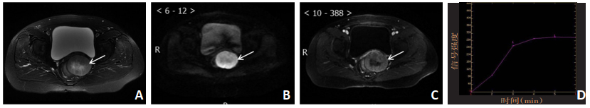
图 1 1例小肠低危险度GISTs典型病例MRI相关征象
A:脂肪抑制T2WI肿瘤呈均匀稍高信号;B:DWI示肿瘤呈高信号,ADC值:1.465×10⁻³ mm2/s;C:增强扫描后肿瘤均匀强化;D:肿瘤时间-信号强化曲线呈流出型

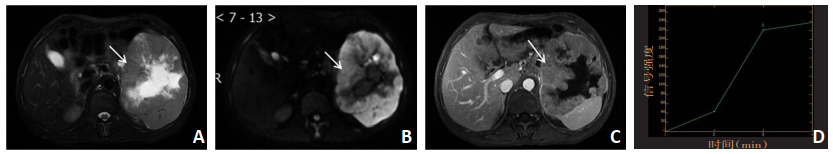
图 2 1例直肠中危险度GISTs典型病例MRI相关征象
A:脂肪抑制T2WI肿瘤呈不均匀高信号;B:DWI示肿瘤呈高信号,ADC值:0.972×10⁻³ mm2/s;C:增强扫描后肿瘤呈不均匀强化;D:肿瘤时间-信号强化曲线呈平台型

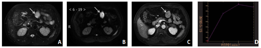
图 3 1例胃高危险度GISTs典型病例MRI相关征象
A:脂肪抑制T2WI肿瘤呈不均匀稍高信号,内可见长T2囊变坏死;B:DWI示肿瘤呈高信号,ADC值:0.613×10⁻³ mm2/s;C:增强扫描后肿瘤呈不均匀强化;D:肿瘤时间-信号强化曲线呈渐增型
表 1 GIST的不同MRI征象及与危险度的相关性

-
[1] Ferrocci G, Rossi C, Bolzon S, et al. Gastrointestinal stromal tumours. Our experience ten years later[J]. Ann Ital Chir, 2011, 82(4):267-272. https://www.ncbi.nlm.nih.gov/pubmed/21834475 [2] Koo DH, Ryu MH, Kim KM, et al. Asian consensus guidelines for the diagnosis and management of gastrointestinal stromal tumor[J]. Cancer Res Treat, 2016, 48(4):1155-1166. doi: 10.4143/crt.2016.187 [3] 赵宇, 刘辉, 习羽, 等.胃间质瘤CT征象与肿瘤危险度分级的关系[J].实用放射学杂志, 2016, 32(6):892-895. doi: 10.3969/j.issn.1002-1671.2016.06.017 [4] Zhou C, Duan X, Zhang X, et al. Predictive features of CT for risk stratifications in patients with primary gastrointestinal stromal tumour[J]. Eur Radiol, 2016, 26(9):3086-3093. doi: 10.1007/s00330-015-4172-7 [5] 李双, 龙学颖, 刘慧.胃间质瘤CT影像特征及纹理参数与危险度分级的相关性[J].中南大学学报, 2019, 44(10):264-270. http://d.old.wanfangdata.com.cn/Periodical/hnykdx201903006 [6] Joensuu H. Risk stratification of patients diagnosed with gastrointestinal stromal tumor[J]. Hum athol, 2008, 39(10):1411-1419. https://www.ncbi.nlm.nih.gov/pubmed/18774375 [7] Sandrasegaran K, Rajesh A, Rydbery J, et al. Gastrointestinal stromal tumors: clinical, radiologic, and pathologic features[J]. AJR Am J Roentgenol, 2005, 184(3):803-811. doi: 10.2214/ajr.184.3.01840803 [8] Yu MH, Lee JM, Baek JH, et al. MRI features of gastrointestinal stromal tumors[J]. AJR Am J Roentgenol, 2014, 203(5):980-989. doi: 10.2214/AJR.13.11667 [9] Miettinen M, Lasota J. Gastrointestinal stromal tumors: pathology and prognosis at different sites[J]. Semin Diagn Pathol, 2006, 23(2):70-83. doi: 10.1053/j.semdp.2006.09.001 [10] Tateishi U, Hasegawa T, Satake M, et al. Gastrointestinal stromal tumor: Correlation of computed tomography findings with tumor grade and mortality[J]. J Comput Assist Tomogr, 2003, 27(5):792-798. doi: 10.1097/00004728-200309000-00018 [11] 高桂花, 夏梦莹, 王淑艳, 等.胃肠道间质瘤危险度分级的磁共振表现与病理对照分析[J].医学影像学杂志, 2017, 27(3):492-496. http://d.old.wanfangdata.com.cn/Periodical/yxyxxzz201703031 [12] Kang TW, Kim SH, Jang KM, et al. Gastrointestinal stromal tumours: correlation of modified NIH risk stratification with diffusion-weightedMR imaging as an imaging biomarker[J]. Eur J Radiol, 2015, 84(1):33- 40. doi: 10.1016/j.ejrad.2014.10.020 [13] Matsui T, Mitsui H, Sekigawa K, et al. A case of aduodenal gastrointestinal stronal tumor diagnosed with the aid of diffusion-weighted magnetic resonance imaging[J]. Clin J Gastroenterol, 2009, 2(10):384- 387. doi: 10.1007/s12328-009-0110-z [14] 李钦海, 陈光.MRI在诊断及评估胃肠道间质瘤侵袭危险度中的应用价值分析[J].现代消化及介入诊疗, 2014, 19(5):320-321. doi: 10.3969/j.issn.1672-2159.2014.05.013 [15] Zhou H, Zhang XM, Zeng NL, et al. Use of conventional MR imaging and diffusion-weighted imaging for evaluating the risk grade of gastrointestinal stromal tumors[J]. J Magn Reson Imaging, 2012, 36(6):1395- 1401. doi: 10.1002/jmri.23784 -

 下载:
下载:
 点击查看大图
图(3) / 表(1)
计量
- 文章访问数: 106
- HTML全文浏览量: 18
- PDF下载量: 5
- 被引次数: 0
