
-
摘要:
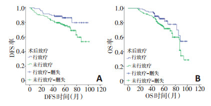
目的 探讨乳腺浸润性微乳头状癌(invasive micropapillary carcinoma,IMPC)的临床病理特征及预后。 方法 回顾性分析2011年1月至2015年12月246例于天津医科大学肿瘤医院收治的乳腺IMPC患者的临床病理资料,分为143例IMPC成分比例> 50%(A组)和103例比例≤50%(B组)两组。多因素分析采用Cox比例风险回归模型,采用Log-rank检验及Kaplan Meier法等进行生存分析。 结果 A组患者的5年无病生存(disease free survival,DFS)和总生存(overall survival,OS)时间均低于B组DFS(76.5% vs.83.6%,P=0.042)和OS(74.1% vs.81.6%,P=0.029)。A组中未行放疗患者的DFS和OS均低于行放疗患者的DFS(χ2=5.219,P=0.022)和OS(χ2=3.963,P=0.047)。Cox比例风险回归模型多因素分析显示,患者人类表皮生长因子受体-2(human epidermal growth factor receptor-2,HER-2)阳性表达(HR=2.989,95% CI:1.400~6.384,P=0.005),乳头侵犯(HR=2.388,95% CI:1.263~ 4.518,P=0.007),4枚以上淋巴结转移(HR=2.076,95% CI:1.080~3.992,P=0.029)为DFS独立危险因素,乳头侵犯(HR=1.951,95% CI:1.054~3.609,P=0.033)为OS独立危险因素,并且乳头侵犯中未行放疗患者的DFS和OS均低于行放疗患者的DFS(χ2=6.541,P=0.011)和OS(χ2=6.455,P=0.012)。 结论 乳腺IMPC作为一种特殊类型乳腺癌,乳头侵犯提示预后较差。IMPC成分比例>50%较比例≤50%乳腺癌患者的预后更差,对于IMPC成分比例>50%或乳头侵犯患者,术后放疗能带来更多生存获益。 Abstract:Objective We aimed to investigate the clinicopathology and prognosis of invasive micropapillary carcinoma (IMPC) of the breast. Methods This was a single-center retrospective study based on the clinicopathological and follow-up data of 246 patients with IMPC who were treated at the Tianjin Medical University Cancer Institute and Hospital between January 2011 and December 2015. The patients were divided into two groups:we included 143 patients with >50% IMPC in group A and the remaining 103 patients in group B. The cox proportional-hazard regression model, Log-rank test, and Kaplan-Meier method were used for analysis. Results The 5-year disease-free survival (DFS; 76.5% vs. 83.6%, P=0.042) and overall survival (OS; 74.1% vs. 81.6%, P=0.029) of group A were lower than those of group B. The DFS (χ2=5.219, P=0.022) and OS (χ2=3.96, P=0.047) of patients who did not receive radiotherapy in group A were lower than those of patients who received radiotherapy. Multivariate Cox regression analysis showed that HER-2 expression (HR=2.989, 95% CI 1.400-6.384, P=0.005), mammilla invasion (HR=2.388, 95% CI 1.263-4.518, P=0.007), and ≥ 4 lymph node metastasis (HR=2.076, 95% CI 1.080-3.992, P=0.029) were independent risk factors for DFS. Mammilla invasion (HR=1.951, 95% CI 1.054-3.609, P=0.033) was an independent risk factor for OS. The DFS (χ2=6.541, P=0.011) and OS (χ2=6.455, P=0.012) in patients with mammilla invasion who did not receive radiotherapy were significantly lower than those of patients who received radiotherapy. Conclusion As a special type of breast cancer, mammilla invasion indicates a poor prognosis. The prognosis of patients with >50% IMPC was worse than that of patients with ≤ 50% IMPC. Postoperative adjuvant radiotherapy may provide survival benefit to patients with IMPC accounting for 50% or mammilla invasion. -
Key words:
- breast carcinoma /
- invasive micropapillary carcinoma /
- prognosis /
- radiotherapy
-
表 1 246例乳腺IMPC患者的单因素分析

表 2 乳腺IMPC患者Cox比例风险回归模型多因素分析

-
[1] Gokce H, Durak MG, Akin MM, et al. Invasive micropapillary carcinoma of the breast:a clinicopathologic study of 103 cases of an unusual and highly aggressive variant of breast carcinoma[J]. Breast J, 2013, 19(4):374-381. http://cn.bing.com/academic/profile?id=b3b986484ca87e19163d3d2a5fbe2be6&encoded=0&v=paper_preview&mkt=zh-cn [2] Carey LA, Perou CM, Livasy CA, et al. Race, breast cancer subtypes, and survival in the Carolina breast cancer study[J]. JAMA, 2006, 295(21):2492-2502. http://cn.bing.com/academic/profile?id=94f3b330f5f7d12e9f12825184ab06fb&encoded=0&v=paper_preview&mkt=zh-cn [3] Wolff AC, Hammond ME, Hicks DG, et al. Recommendations for human epidermal growth factor receptor 2 testing in breast cancer:American society of clinical oncology/college of American pathologists clinical practice guideline update[J]. J Clin Oncol, 2013, 31(31):3997-4013. http://cn.bing.com/academic/profile?id=688ede51addb8eb8b7500f0922df8aa3&encoded=0&v=paper_preview&mkt=zh-cn [4] Hammond ME, Hayes DF, Wolff AC, et al. American society of clinical oncology/college of American pathologists guideline recommendations for immunohistochemical testing of estrogen and progesterone receptors in breast cancer[J]. J Oncol Pract, 2010, 6(4):195-197. http://cn.bing.com/academic/profile?id=ecd3a78d65991e462bac75359ec5e562&encoded=0&v=paper_preview&mkt=zh-cn [5] Sauter G, Lee J, Bartlett JM, et al. Guidelines for human epidermal growth factor receptor 2 testing:biologic and methodologic considerations[J]. J Clin Oncol, 2009, 27(8):1323-1333. http://cn.bing.com/academic/profile?id=78367321ae7a753620b6e5d1ce383265&encoded=0&v=paper_preview&mkt=zh-cn [6] Chen AC, Paulino AC, Schwartz MR, et al. Population-based comparison of prognostic factors in invasive micropapillary and invasive ductal carcinoma of the breast[J]. Br J Cancer, 2014, 111(3):619-622. http://www.wanfangdata.com.cn/details/detail.do?_type=perio&id=f7a6c3e5521d4c5d7424a71ce04a3cdb [7] Yamaguchi R, Tanaka M, Kondo K, et al. Characteristic morphology of invasive micropapillary carcinoma of the breast:an immunohistochemical analysis[J]. Jpn J Clin Oncol, 2010, 40(8):781-787. http://cn.bing.com/academic/profile?id=6e6d55d0d18c312de2fed64e49396b95&encoded=0&v=paper_preview&mkt=zh-cn [8] Kim MJ, Gong G, Joo HJ, et al. Immunohistochemical and clinicopathologic characteristics of invasive ductal carcinoma of breast with micropapillary carcinoma component[J]. Arch Pathol Lab Med, 2005, 129(10):1277-1282. http://cn.bing.com/academic/profile?id=f9f7bcc3376e369fcf7ad945916b59f2&encoded=0&v=paper_preview&mkt=zh-cn [9] Marchiò C, Iravani M, Natrajan R, et al. Mixed micropapillary-ductal carcinomas of the breast:a genomic and immunohistochemical analysis of morphologically distinct components[J]. J Pathol, 2009, 218(3):301-315. http://cn.bing.com/academic/profile?id=37a094718d17b1a20f741e8b81247e6d&encoded=0&v=paper_preview&mkt=zh-cn [10] Fan Y, Lang RG, Wang Y, et al. Relationship between expression of cell adhesion molecules and metastatic potential in invasive micropapillary carcinoma of breast[J]. Zhonghua Bing Li Xue Za Zhi, 2004, 33(4):308-311. http://cn.bing.com/academic/profile?id=e8d838031ab937acf58ec9cbf0cd1137&encoded=0&v=paper_preview&mkt=zh-cn [11] Meng F, Liu B, Xie G, et al. Amplification and overexpression of PSCA at 8q24 in invasive micropapillary carcinoma of breast[J]. Breast Cancer Res Treat, 2017, 166(2):383-392. http://www.wanfangdata.com.cn/details/detail.do?_type=perio&id=8f069a5ed7336186d810dbe0db966955 [12] Yang YL, Liu BB, Zhang X, et al. Invasive micropapillary carcinoma of the breast:an update[J]. Arch Pathol Lab Med, 2016, 140(8):799-805. http://cn.bing.com/academic/profile?id=4d9ce4a850436188ff8272654f1a50fd&encoded=0&v=paper_preview&mkt=zh-cn [13] Haffty BG, Mahmoud O. The evolution of regional nodal irradiation in breast cancer[J]. Breast J, 2015, 21(1):32-41. http://cn.bing.com/academic/profile?id=f255a140a7d65d34fa53e260ea45e485&encoded=0&v=paper_preview&mkt=zh-cn [14] Wu SG, Zhang WW, Sun JY, et al. Postoperative radiotherapy for invasive micropapillary carcinoma of the breast:an analysis of surveillance, epidemiology, and end results database[J]. Cancer Manag Res, 2017, 9:453-459. http://cn.bing.com/academic/profile?id=8c95b15ea4bbef6ff1aba649201ee3a8&encoded=0&v=paper_preview&mkt=zh-cn [15] Lewis GD, Xing Y, Haque W, et al. Prognosis of lymphotropic invasive micropapillary breast carcinoma analyzed by using data from the national cancer database[J]. Cancer Commun (Lond), 2019, 39(1):60. http://cn.bing.com/academic/profile?id=05f834b281cff6e05f9add258477545c&encoded=0&v=paper_preview&mkt=zh-cn [16] Cui ZQ, Feng JH, Zhao YJ. Clinicopathological features of invasive micropapillary carcinoma of the breast[J]. Oncol Lett, 2015, 9(3):1163-1166. http://d.old.wanfangdata.com.cn/Periodical/nfmwk201906014 [17] Chen AC, Paulino AC, Schwartz MR, et al. Prognostic markers for invasive micropapillary carcinoma of the breast:a population-based analysis[J]. Clin Breast Cancer, 2013, 13(2):133-139. http://cn.bing.com/academic/profile?id=c282e25cdf0e2fd44badc0ded0b49653&encoded=0&v=paper_preview&mkt=zh-cn -

 下载:
下载:
 点击查看大图

图(2) / 表(2)
计量
- 文章访问数: 460
- HTML全文浏览量: 260
- PDF下载量: 15
- 被引次数: 0
