
Prediction of complete pathological response to neoadjuvant chemotherapy in patients with breast cancer with reduced microcalcification
-
摘要:
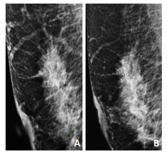
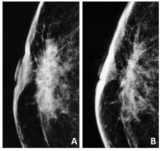
目的 探讨乳腺癌患者新辅助化疗(neoadjuvant chemotherapy,NAC)后影响微钙化(mcrocalcification,MC)改变的因素及MC减少与肿瘤病理完全反应(pathological complete response,pCR)的相关性。 方法 收集2015年1月至2018年12月天津医科大学肿瘤医院215例乳腺癌患者的临床资料,分为范围改变组及数量改变组,评估影响MC改变的因素。根据MC是否减少进行分组,分为MC范围缩小组及MC数量减少组,分析不同分子分型中MC减少与pCR的相关性。采用受试者工作特征曲线(receiver operat-ing characteristic curve,ROC)评价乳腺X线摄影(mammography,MG)检查中MC减少对pCR敏感性、特异性的预测。 结果 MC呈弥散分布,范围>2 cm,数量>20个患者更易发生MC减少。MC范围缩小组较非缩小组易发生pCR。MC数量减少组与非减少组比较差异无统计学意义,分子分型不是MC范围缩小及数量减少与pCR的影响因素。MC范围缩小组预测pCR的敏感度为77.78%、特异度为57.45%(P=0.000 1)。 结论 乳腺癌患者NAC后MC改变因素为MC范围、数量及分布,MC范围缩小患者的pCR率高,但MC范围缩小对预测pCR准确性较低,暂不推荐MG检查评估NAC后是否达到pCR。 Abstract:Objective To investigate causes and factors affecting microcalcification (MC) changes after neoadjuvant chemotherapy (NAC) in patients with breast cancer and to assess the correlation between MC reduction and complete remission rate [pathological complete response (pCR)] of tumors. Methods Clinical data of 215 patients with breast cancer who visited Tianjin Medical University Cancer Hospital from January 1, 2015 to December 31, 2018 were collected. The patients were grouped according to MC range and number of changes, and factors that affected MC changes were evaluated. According to whether MC decreased or not, the patient group was divided into the MC range and MC number reduction groups, and the correlation between the decrease in MC and pCR, assessed using different molecular typing methods, was analyzed. The sensitivity and specificity of the receiver operating characteristic curve analysis (ROC) were used to evaluate the accuracy of MC reduction in predicting pCR. Results The patients with a distribution of diffuse, initial MC range of >2 cm and MC quantity of >20 were more likely to have an MC reduction. The pCR rate was higher in the group with reduced and non-reduced MC. No significant difference was found between the group with decreased MC and the control group. The decreases in MC according to the different molecular typing methods were independent of factors affecting pCR. Reduction in MC range predicts pCR with a sensitivity of 77.78 and specificity of 57.45 (P=0.0001). Conclusions The change factors of MC in patients with breast cancer after neoadjuvant chemotherapy were the range, number, and distribution of calcification. The pCR rate of the patients with reduced MC was high, but the accuracy of pCR prediction based on reduced MG was low. Thus, mammography was not recommended for evaluating pCR after neoadjuvant chemotherapy. -
表 1 215例乳腺癌患者行NAC后MC的病理特征和MC变化模式

表 2 MC减少与pCR的相关性分析

-
[1] Colomer R, Saura C, Sánchez-Rovira P, et al. Neoadjuvant management of early breast cancer:a clinical and investigational position statement[J]. Oncologist, 2019, 24(5):603-611. doi: 10.1634/theoncologist.2018-0228 [2] 中国乳腺癌新辅助治疗专家组.中国乳腺癌新辅助治疗专家共识(2019年版)[J].中国癌症杂志, 2019, 29(5):390-400. http://www.wanfangdata.com.cn/details/detail.do?_type=perio&id=zgazzz201905009 [3] Panato C, Abusamaan K, Bidoli E, et al. Survival after the diagnosis of breast or colorectal cancer in the GAZA strip from 2005 to 2014[J]. BMC Cancer, 2018, 18(1):632. doi: 10.1186/s12885-018-4552-x [4] Sener SF, Sargent RE, Lee C, et al. MRI does not predict pathologic complete response after neoadjuvant chemotherapy for breast cancer[J]. J Surg Oncol, 2019, 120(6):903-910. http://www.wanfangdata.com.cn/details/detail.do?_type=perio&id=0cd7641fae01f6e0b7288acca8997423 [5] Qi X, Chen A, Zhang P, et al. Mammographic calcification can predict outcome in women with breast cancer treated with breast-conserving surgery[J]. Oncol Lett, 2017, 14(1):79-88. doi: 10.3892/ol.2017.6112 [6] Koning JL, Davenport KP, Poole PS, et al. Breast imaging-reporting and data system (BI-RADS) classification in 51 excised palpable pediatric breast masses[J]. J Pediatr Surg, 2015, 50(10):1746-1750. doi: 10.1016/j.jpedsurg.2015.02.062 [7] Kim YS, Chang JM, Moon HG, et al. Residual mammographic microcalcifications and enhancing lesions on MRI after neoadjuvant systemic chemotherapy for locally advanced breast cancer:correlation with histopathologic residual tumor size[J]. Ann Surg Oncol, 2016, 23(4):1135-1142. doi: 10.1245/s10434-015-4993-2 [8] An YY, Kim SH, Kang BJ. Residual microcalcifications after neoadjuvant chemotherapy for locally advanced breast cancer:comparison of the accuracies of mammography and MRI in predicting pathological residual tumor[J]. World J Surg Oncol, 2017, 15(1):198. doi: 10.1186/s12957-017-1263-8 [9] Gu YL, Pan SM, Ren J, et al. Role of magnetic resonance imaging in detection of pathologic complete remission in breast cancer patients treated with neoadjuvant chemotherapy:a Meta-analysis[J].Clin Breast Cancer, 2017, 17(4):245-255. doi: 10.1016/j.clbc.2016.12.010 [10] Feliciano Y, Mamtani A, Morrow M, et al. Do calcifications seen on mammography after neoadjuvant chemotherapy for breast cancer always need to be excised[J]?Ann Surg Oncol, 2017, 24(6):1492-1498. doi: 10.1245/s10434-016-5741-y [11] Golan O, Amitai Y, Menes T. Does change in microcalcifications with neoadjuvant treatment correlate with pathological tumour response[J]?Clin Radiol, 2016, 71(5):458-463. http://cn.bing.com/academic/profile?id=7ffce276813032158d8c289312372451&encoded=0&v=paper_preview&mkt=zh-cn [12] Adrada BE, Huo L, Lane DL, et al. Histopathologic correlation of residual mammographic microcalcifications after neoadjuvant chemotherapy for locally advanced breast cancer[J]. Ann Surg Oncol, 2015, 22(4):1111-1117. doi: 10.1245/s10434-014-4113-8 [13] Li JJ, Chen C, Gu Y, et al. The role of mammographic calcification in the neoadjuvant therapy of breast cancer imaging evaluation[J].PLoS One, 2014, 9(2):e88853. doi: 10.1371/journal.pone.0088853 [14] Fushimi A, Kudo R, Takeyama H. Do decreased breast microcalcifications after neoadjuvant chemotherapy predict pathologic complete response[J]?Clin Breast Cancer, 2020, 20(1):e82-e88. http://cn.bing.com/academic/profile?id=6b55a96cad1a751953f2d89a083d528b&encoded=0&v=paper_preview&mkt=zh-cn [15] Yim H, Ha T, Kang DK, et al. Change in microcalcifications on mammography after neoadjuvant chemotherapy in breast cancer patients:correlation with tumor response grade and comparison with lesion extent[J]. Acta Radiol, 2019, 60(2):131-139. doi: 10.1177/0284185118776491 -

 下载:
下载:
 点击查看大图
图(2) / 表(2)
计量
- 文章访问数: 80
- HTML全文浏览量: 33
- PDF下载量: 5
- 被引次数: 0
