
Clinicopathological characteristics of 83 lymph node-negative rectal neuroendocrine tumors
-
摘要:
目的 探讨淋巴结阴性直肠神经内分泌肿瘤(rectal neuroendocrine neoplasms,R-NENs)的临床病理特征。 方法 回顾性分析2012年12月至2019年12月中日友好医院83例淋巴结阴性R-NENs患者的临床病理资料,并定期随访。 结果 83例患者中男性49例(59.0%)、女性34例(41.0%);平均年龄(43.3±11.4)岁;61例(75.5%)患者主要因非特异性症状就诊;肿瘤单发75例(90.4%);肿瘤平均直径为(0.8±0.7)cm;主要浸润黏膜层及黏膜下层80例(96.4%);病理分级以G1为主,共65例(78.3%),Ki-67指数平均值为(2.1±1.7)%;肿瘤分期Ⅰ期78例(94.0%)。免疫组织化学法检测CgA阳性29例(34.9%)。治疗方式使用内镜下切除67例(80.7%),手术16例(19.3%)。中位随访时间24(3~90)个月,5年生存率100%,2例(2.4%)复发。肿瘤复发与Ki-67阳性指数具有显著相关性(P=0.025);肿瘤浸润深度与肿瘤直径具有相关性(P=0.030)。Kaplan-Meier法分析得出治疗方式、肿瘤分级对预后复发的差异具有统计学意义(P=0.031、0.001)。 结论 淋巴结阴性R-NENs直径>1.0 cm相对容易浸及固有肌层,直径≤ 1.0 cm也有浸及固有肌层的可能,建议此类患者行超声内镜(EUS)检查以决定治疗方式。内镜下切除为淋巴结阴性R-NENs的主要治疗方式,Ki-67指数较高患者治疗后相对容易复发。 Abstract:Objective To explore the clinicopathological characteristics of lymph node-negative rectal neuroendocrine neoplasms (RNENs). Methods We retrospectively analyzed and regularly followed up the clinical and pathological data of 83 patients with lymph node-negative rectal NENs treated at China-Japan Friendship Hospital between December 2012 to December 2019. Results Among the 83 patients, 49 (59%) were male and 34 (41%) were female with an average age of (43.3±11.4) years. Of the patients, 61 (75.5%) were mainly treated for nonspecific symptoms, 75 (90.4%) had a single tumor with an average size of (0.8±0.7) cm, 80 (96.4%) showed tumor infiltration into the mucosal and submucosal layers, and 65 (78.3%) predominantly had tumors of pathological grade G1. The average Ki-67 index was (2.1±1.7)%, with 78 (94%) patients having stage I tumors. Twenty-nine (34.9%) patients showed CgA positivity on immunohistochemical analysis. As for treatment, 67 (80.7%) patients underwent endoscopic resection, and 16 (19.3%) patients underwent surgery. The median follow-up time was 24 (3-90) months, with a 100% 5-year survival rate and relapse in 2 (2.4%) patients. Tumor recurrence was significantly correlated with the Ki-67 positive index (P=0.025), and tumor infiltration depth was correlated with the tumor diameter (P=0.03). Kaplan-Meier analysis showed that different treatment mode and tumor grade on prognosis and recurrence was statistically significant (Log-rank P=0.031, 0.001). Conclusion Lymph node-negative rectal neuroendocrine neoplasms with a diameter >1 cm infiltrate the muscularis propria relatively easily and those ≤ 1 cm may also infiltrate the muscularis propria. It is recommended that all patients undergo ultrasound enteroscope (EUS) to determine the treatment choice. Endoscopic resection is the main treatment for lymph node-negative rectal neuroendocrine neoplasms. Patients with a high Ki-67 index are relatively prone to relapse after treatment. -

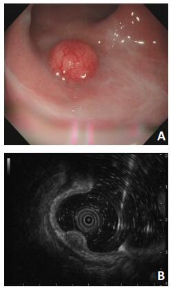
图 1 R-NENs超声内镜下表现
A:电子肠镜:距肛门5 cm处可见一直径约1.0 cm的隆起病变,表面尚光滑;B:超声内镜:病变位于黏膜及黏膜肌层的稍低回声结节,大小0.7 cm×0.5 cm,黏膜下层及固有肌层完整连续

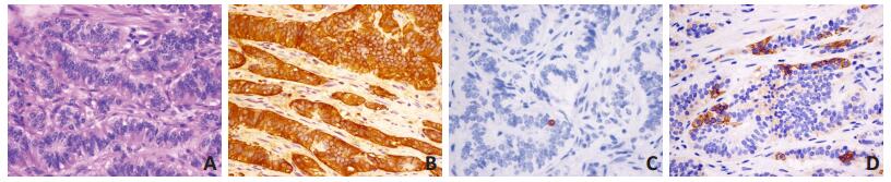
图 2 R-NENs的病理学特征
A:1例淋巴结阴性R-NENs G1患者(H&E×400),可见肿瘤细胞排列呈梁索状;B:免疫组织化学染色肿瘤细胞CgA阳性(IHC×400);C:免疫组织化学染色肿瘤细胞Ki-67为1%(IHC×400);D:免疫组织化学染色肿瘤细胞SSTR2阳性(IHC×400)
表 1 83例淋巴结阴性R-NENs患者临床病理特征

-
[1] Dasari A, Shen C, Halperin D, et al. Trends in the incidence, prevalence, and survival outcomes in patients with neuroendocrine tumors in the united states[J]. JAMA Oncol, 2017, 3(10):1335-1342. http://cn.bing.com/academic/profile?id=b862c878d2fe15533ba67fb06e82bf56&encoded=0&v=paper_preview&mkt=zh-cn [2] Tsai HJ, Wu CC, Tsai CR, et al. The epidemiology of neuroendocrine tumors in taiwan:a nation-wide cancer registry-based study[J]. PLoS One, 2013, 22(8):e62487. https://pubmed.ncbi.nlm.nih.gov/23614051/ [3] Cho MY, Kim JM, Sohn JH, et al. Current trends of the incidence and pathological diagnosis of gastroenteropancreatic neuroendocrine tumors (GEP-NETs) in Korea 2000-2009:multicenter study[J]. Cancer Res Treat, 2012, 44(3):157-165. http://d.old.wanfangdata.com.cn/OAPaper/oai_pubmedcentral.nih.gov_3467418 [4] Ito T, Igarashi H, Nakamura K, et al. Epidemiological trends of pancreatic and gastrointestinal neuroendocrine tumors in Japan:a nationwide survey analysis[J]. J Gastroenterol, 2015, 50(1):58-64. http://cn.bing.com/academic/profile?id=8b4ac966cc1ecb2cddf5fa227958a39b&encoded=0&v=paper_preview&mkt=zh-cn [5] 邱旭东, 刘猛, 刘青, 等.903例神经内分泌肿瘤发病部位与病理特征分析[J].中华胃肠外科杂志, 2017, 20(9):993-996. http://d.old.wanfangdata.com.cn/Periodical/zgwcwkzz201709013 [6] Bosman FT, Carneiro F, Hruban RH, et al. WHO classification of tumours of the digestive system[M]. Lyon:IARC, 2010. [7] Edge SB, Byrd DR, Compton CC, et al. AJCC Cancer Staging Manual(ed 7th)[M]. New York:Springer, 2010. [8] Moon CM, Huh KC, Jung SA, et al. Long-term clinical outcomes of rectal neuroendocrine tumors according to the pathologic status after initial endoscopic resection:a kasid multicenter study[J]. Am J Gastroenterol, 2016, 111(9):1276-1285. http://europepmc.org/abstract/MED/27377520 [9] 黄玉庭, 贾茹, 徐倩, 等.不同分期结肠与直肠神经内分泌肿瘤的预后分析[J].中华肿瘤杂志, 2019, 41(2):146-151. http://d.old.wanfangdata.com.cn/Periodical/zhzl201902014 [10] Moon SH, Hwang JH, Sohn DK, et al. Endoscopic submucosal dissection for rectal neuroendocrine (carcinoid) tumors[J]. J Laparoendosc Adv Surg Tech A, 2011, 21(8):695-699. http://cn.bing.com/academic/profile?id=34d234495e4957688508ed1cf55a8c0a&encoded=0&v=paper_preview&mkt=zh-cn [11] Kim HR, Lee WY, Jung KU, et al. Transanal endoscopic microsurgery for the treatment of well-differentiated rectal neuroendocrine tumors[J]. J Korean Soc Coloproctol, 2012, 28(4):201-204. http://d.old.wanfangdata.com.cn/OAPaper/oai_pubmedcentral.nih.gov_3440489 [12] Mc Connell, Yarrow J. Surgical management of rectal carcinoids:trends and outcomes from the Surveillance, Epidemiology, and End Results database (1988 to 2012)[J]. Am J Surg, 2016, 211(5):877-885. http://cn.bing.com/academic/profile?id=4e64e909d06d69a3b7502e2397f4c717&encoded=0&v=paper_preview&mkt=zh-cn [13] Sohn B, Kwon Y, Ryoo SB, et al. Predictive factors for lymph node metastasis and prognostic factors for survival in rectal neuroendocrine tumors[J]. J Gastrointest Surg, 2017, 21(12):2066-2074. http://www.wanfangdata.com.cn/details/detail.do?_type=perio&id=224e864eb454b4644c8a9ba1b18fd9bd [14] Folkert IW, Sinnamon AJ, Concors SJ, et al. Grade is a dominant risk factor for metastasis in patients with rectal neuroendocrine tumors[J]. Ann Surg Oncol, 2020, 27(3):855-863. http://cn.bing.com/academic/profile?id=7ad6ea8cd0af0772960867fcd04b0855&encoded=0&v=paper_preview&mkt=zh-cn [15] Motohiro K, Koji I, Norio S, et al. Neuroendocrine tumors of the large intestine:clinicopathological features and predictive factors of lymph node metastasis[J]. Front Oncol, 2016, 18(6):173. http://cn.bing.com/academic/profile?id=c011b7b7a5038011866bbec8ec131c80&encoded=0&v=paper_preview&mkt=zh-cn [16] Caplin M, Sundin A, Nillson O, et al. ENETS consensus guidelines for the management of patients with digestive neuroendocrine neoplasms:colorectal neuroendocrine neoplasms[J]. Neuroendocrinology, 2012, 95(2):98-119. http://cn.bing.com/academic/profile?id=be970659419af6c6580f8be4c1a65bdd&encoded=0&v=paper_preview&mkt=zh-cn [17] Gu Q, Lin YM, Cen L, et al. Endoscopic ultrasonography is useful in the diagnosis and treatment of rectal neuroendocrine neoplasms:a case series[J]. J Zhejiang Univ Sci B, 2019, 20(10):861-864. http://d.old.wanfangdata.com.cn/Periodical/zjdxxbb-e201910009 [18] Tsukamoto S, Fujita S, Yamaguchi T, et al. Clinicopathological characteristics and prognosis of rectal well-differentiated neuroendocrine tumors[J]. Int J Colorectal Dis, 2008, 23(11):1109-1113. http://www.wanfangdata.com.cn/details/detail.do?_type=perio&id=84550c96317bc105a0983f50e111f79d [19] Chablaney S, Zator ZA, Kumta NA. Diagnosis and management of rectal neuroendocrine tumors[J]. Clin Endosc, 2017, 50(6):530-536. http://cn.bing.com/academic/profile?id=ce30f75469046363abed586574724adf&encoded=0&v=paper_preview&mkt=zh-cn [20] Kulke M, Shah MH, Benson AB, et al. Neuroendocrine Tumors, Version 1.2015[J]. J Natl Compr Canc Netw, 2015, 13(1):78-108. http://cn.bing.com/academic/profile?id=3c735aec2319abbd2e63a2800b4573a0&encoded=0&v=paper_preview&mkt=zh-cn [21] Joon JH, Cheung DY, Lee SJ, et al. Endoscopic resection yields reliable outcomes for small rectal neuroendocrine tumors[J]. Dig Endosc, 2014, 26(4):556-563. http://cn.bing.com/academic/profile?id=0d53f383153109175fc7003e5f6bd7a6&encoded=0&v=paper_preview&mkt=zh-cn [22] Sung HY, Kim SW, Kang WK, et al. Long-term prognosis of an endoscopically treated rectal neuroendocrine tumor[J]. Eur J Gastroenterol Hepatol, 2012, 24(8):978-983. http://cn.bing.com/academic/profile?id=738881bc698e0e4bbe1575e5dae464a8&encoded=0&v=paper_preview&mkt=zh-cn -

 下载:
下载:


 点击查看大图
图(5) / 表(1)
计量
- 文章访问数: 104
- HTML全文浏览量: 8
- PDF下载量: 5
- 被引次数: 0
