
Analysis of the outcomes of esophageal precancerous lesions between 2006 and 2016 in Feicheng City, a high-incidence area of esophageal cancer
-
摘要:
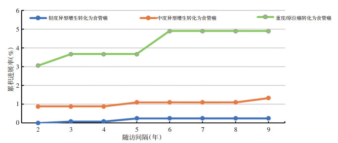
目的 研究食管癌高发区中国山东省肥城市食管癌前病变自然转归情况,为食管癌前病变及食管鳞癌的防治工作提供科学依据。 方法 回顾性收集山东省肥城市2006年至2016年期间进行内镜病理诊断且未治疗并进行二次内镜随访的受检者资料进行分析,描述癌前病变病例的具体复查结果,计算进展病例的累积进展率和进展时间,分析食管癌前病变病例的转归情况。 结果 本研究共纳入1 834例食管癌前病变病例,其中1 148例(62.6%)癌前病变发生逆转,148例(8.1%)发生进展。逆转为正常状态的病例共234例(12.8%),进展为食管癌共17例(0.9%)。各级别癌前病变进展为食管癌的病例比例由高至低依次为:重度异型增生/原位癌(4.9%)、中度异型增生(1.3%)、轻度异型增生(0.2%);其发生癌变的中位进展时间由高至低依次为:轻度异型增生(5.62年)、中度异型增生(1.76年)和重度异型增生/原位癌(1.61年)。轻度异型增生9年累积进展为重度异型增生/原位癌及以上的进展率远小于中度异型增生(1.81% vs.9.98%),重度异型增生/原位癌进展为食管癌的累积进展率始终高于中度和轻度异型增生。 结论 超过一半以上的癌前病变会逆转为较低级别病变或正常状态;食管癌前病变的累积癌变率随病变级别的增高而增大,中位进展时间随病变级别的增高而缩短。大多数癌前病变进展为食管癌的时间间隔基本与《癌症早诊早治上消化道癌筛查及早诊早治技术方案》中的随访间隔相符,可适当对轻度异型增生患者的随访间隔缩短为每2年1次。 Abstract:Objective Studying the prognosis of esophageal precancerous lesions in Feicheng City and to provide scientific basis for the prevention and control of esophageal squamous cell carcinomas. Methods Retrospective analyze the data of subjects who were not given any medical treatment after endoscopic pathological diagnosis and accepted secondary endoscopic follow-up screening in oFeicheng City from 2006-2016. Describe the results of reexaminations and calculate the cumulative progression rate and progression time to analysis the outcomes of esophageal precancerous lesions. Results A total of 1, 834 cases of precancerous esophageal lesions were included in our study, of which 1, 148 (62.6%) were reversed and 148 (8.1%) were advanced. A total of 234 (12.8%) cases were reversed to normal state, and 17 (0.9%)cases progressed to esophageal cancer. The proportions of precancerous lesions progressing to esophageal cancer from high to low are: severe dysplasia/carcinoma in situ (4.9%), moderate dysplasia (1.3%), mild dysplasia (0.2%); and the median times of cancerous changes from high to low are: mild dysplasia (5.62 years), moderate dysplasia (1.76 years) and severe dysplasia/carcinoma in situ (1.61 years). The 9-year cumulative progression of mild dysplasia to severe dysplasia/carcinoma in situ and above is much less than moderate dysplasia (1.81% vs. 9.98%). The cumulative carcinoma progression rate of severe dysplasia/carcinoma in situ was consistently higher than that of moderate and mild esophageal carcinoma. Conclusions More than half of the precancerous lesions will be reversed to normal or lower-grade lesions. The cumulative carcinogenesis rate of esophageal precancerous lesions increased with the grade of the lesion, and the median time to progression decreased with increasing lesion grade. The time interval for most precancerous lesions to progress to esophageal cancer was roughly consistent with the follow-up interval in the technical plan for the early diagnosis and treatment of cancer. The follow-up frequency for patients with mild dysplasia should be reduced to once every two years. -
Key words:
- esophageal squamous cell carcinoma /
- precancerous lesions /
- outcomes /
- screening interval
-
表 1 筛查前研究对象一般人口学特征

表 2 食管癌前病变转归情况

表 3 食管癌前病变进展规律

-
[1] Bray F, Ferlay J, Soerjomataram I, et al. Global cancer statistics 2018:GLOBOCAN estimates of incidence and mortality worldwide for 36 cancers in 185 countries[J]. CA Cancer J Clin, 2018, 68(6):394-424. doi: 10.3322/caac.21492 [2] 陈茹, 郑荣寿, 张思维, 等.2015年中国食管癌发病和死亡情况分析[J].中华预防医学杂志, 2019, 53(11):1094-1097. [3] Arnold M, Soerjomataram I, Ferlay J, et al. Global incidence of oesophageal cancer by histological subtype in 2012[J]. Gut, 2015, 64(3):381-387. doi: 10.1136/gutjnl-2014-308124 [4] 连士勇, 刘志才, 程兰平, 等.林州市1959~2002年食管癌贲门癌患者的生存分析[J].中国肿瘤, 2007, 16(2):77-78. doi: 10.3969/j.issn.1004-0242.2007.02.003 [5] Wei WQ, Hao CQ, Guan CT, et al. Esophageal histological precursor lesions and subsequent 8.5-year cancer risk in a population-based prospective study in China[J]. Am J Gastroenterol, 2020, 115(7):1036-1044. http://journals.lww.com/ajg/Abstract/2020/07000/Esophageal_Histological_Precursor_Lesions_and.17.aspx [6] 刘曙正, 于亮, 李变云, 等.林州市食管癌筛查病例与非筛查病例生存情况比较[J].中华预防医学杂志, 2018, 52(3):238-242. doi: 10.3760/cma.j.issn.0253-9624.2018.03.005 [7] 张楠, 马恒敏, 孙雅文, 等.山东省2013-2016年农村居民食管癌社会性筛查结果分析[J].中华肿瘤防治杂志, 2017, 24(5):287-290. [8] 王贵齐, 魏文强, 主编.上消化道癌筛查及早诊早治技术方案:2020年试行版[M].北京:人民卫生出版社, 2020:63-139. [9] 马丹, 杨帆, 廖专, 等.中国早期食管癌筛查及内镜诊治专家共识意见(2014年, 北京)[J].中国实用内科杂志, 2015, 35(4):320-337. [10] 沈琼, 王东煜, 项芸岩, 等.食管癌前增生营养阻断研究初步报告[J].中国肿瘤临床, 1991, 18(1):32-34. [11] Taylor PR, Abnet CC, Dawsey SM. Squamous dysplasia——the precursor lesion for esophageal squamous cell carcinoma[J]. Cancer Epidemiol Biomarkers Prev, 2013, 22(4):540-552. doi: 10.1158/1055-9965.EPI-12-1347 [12] 张志镒, 吴正奇, 卢林芝, 等.2009-2017年武威市凉州区上消化道癌筛查和随访结果分析[J].中华肿瘤防治杂志, 2019, 26(23):1750-1755. [13] 何波.食管贲门癌前病变转归及早期癌的内镜治疗随访分析[D].河北医科大学, 2015: 1-41. [14] 王士杰, 张立玮, 温登瑰, 等.食管及贲门癌前病变自然史内镜筛查分析[J].中国肿瘤临床, 2007, 34(7):370-373. doi: 10.3969/j.issn.1000-8179.2007.07.003 [15] Wang JW, Guan CT, Wang LL, et al. Natural history analysis of 101 severe dysplasia and esophageal carcinoma cases by endoscopy[J]. Gastroenterol Res Pract, 2017, 2017:9612854. [16] Dawsey SM, Lewin KJ, Wang GQ, et al. Squamous esophageal histology and subsequent risk of squamous cell carcinoma of the esophagus. A prospective follow-up study from Linxian, China[J]. Cancer, 1994, 74(6):1686-1692. doi: 10.1002/1097-0142(19940915)74:6<1686::AID-CNCR2820740608>3.0.CO;2-V [17] Wang GQ, Abnet CC, Shen Q, et al. Histological precursors of oesophageal squamous cell carcinoma:results from a 13 year prospective follow up study in a high risk population[J]. Gut, 2005, 54(2):187-192. doi: 10.1136/gut.2004.046631 -

 下载:
下载:
 点击查看大图
图(1) / 表(3)
计量
- 文章访问数: 240
- HTML全文浏览量: 99
- PDF下载量: 41
- 被引次数: 0
