
Impact of prostate-specific membrane antigen positron emission tomography/computed tomography on initial tumor-node-metastasis staging and treatment strategy for intermediate-and high-risk prostate cancers
-
摘要:
目的 评估前列腺特异性膜抗原(prostate-specific membrane antigen,PSMA)正电子发射计算机断层显像(positron emission tomography/computed tomography,PET/CT)对中高危前列腺癌初始临床分期及治疗策略的影响。 方法 回顾性分析2019年10月至2021年11月63例于重庆大学附属肿瘤医院初诊的中高危前列腺癌患者的临床及影像资料。比较PSMA PET/CT和传统影像学对前列腺癌病灶检出率及TNM分期的差异,评估PSMA PET/CT对治疗计划的影响。 结果 PSMA PET/CT对区域外淋巴结和骨转移的检出率分别为23.81%(15/63)和52.38%(33/63),较传统影像学的检出率3.17%(2/63)和25.40%(16/63)高,差异具有统计学意义(均P<0.001)。PSMA PET/CT检查后53.97%(34/63)患者TNM分期发生改变,N、M分期上调(均P<0.05);22.22%(14/63)患者的治疗计划发生改变。 结论 PSMA PET/CT检查后上调中高危前列腺癌初始TNM分期中的N、M分期,导致超过20%患者临床治疗策略发生改变。PSMA PET/CT可成为精准评估中高危前列腺癌肿瘤负荷的首选影像学检查。 -
关键词:
- 前列腺特异性膜抗原 /
- 前列腺癌 /
- 正电子发射断层显像术
Abstract: Objective : To evaluate the effect of prostate-specific membrane antigen (PSMA) positron emission tomography/computed tomography (PET/CT) on primary clinical tumor-node-metastasis (TNM) staging and treatment strategies for intermediate-and high-risk prostate cancers. Methods: The clinical and imaging data of 63 patients with intermediate-and high-risk prostate cancers at Chongqing University Cancer Hospital between October 2019 and November 2021 were retrospectively evaluated. The differences between PSMA PET/CT and conventional imaging in lesion detection rate and initial prostate cancer TNM stage were analyzed, and the effect of PSMA PET/CT on treatment strategies was assessed. Results: The detection rates of lymph node and bone metastases using PSMA PET/CT were 23.81% (15/63) and 52.38% (33/63), respectively, 3.17% (2/63) and 25.40% (16/63) more than those determined using traditional imaging, and the differences were significant (all P<0.001). The TNM stage changed for 53.97% (34/63) patients after PSMA PET/CT examination, and the N and M stages were upregulated (all P<0.05). A total of 22.22% (14/63) patients had changes in the treatment plan. Conclusions: The initial N and M stages of intermediate-and high-risk prostate cancers had been upregulated after PSMA PET/CT examination, which led to a change in clinical treatment strategies for more than 20% of patients. PSMA PET/CT may potentially be the first-line imaging modality to accurately assess the whole-body tumor burden in patients with intermediate-and high-risk prostate cancers. -
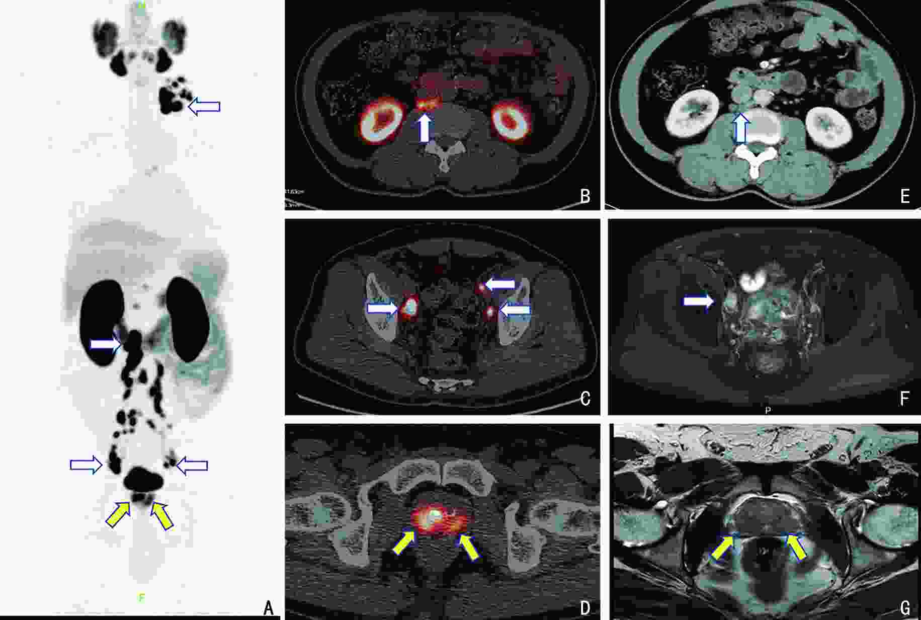
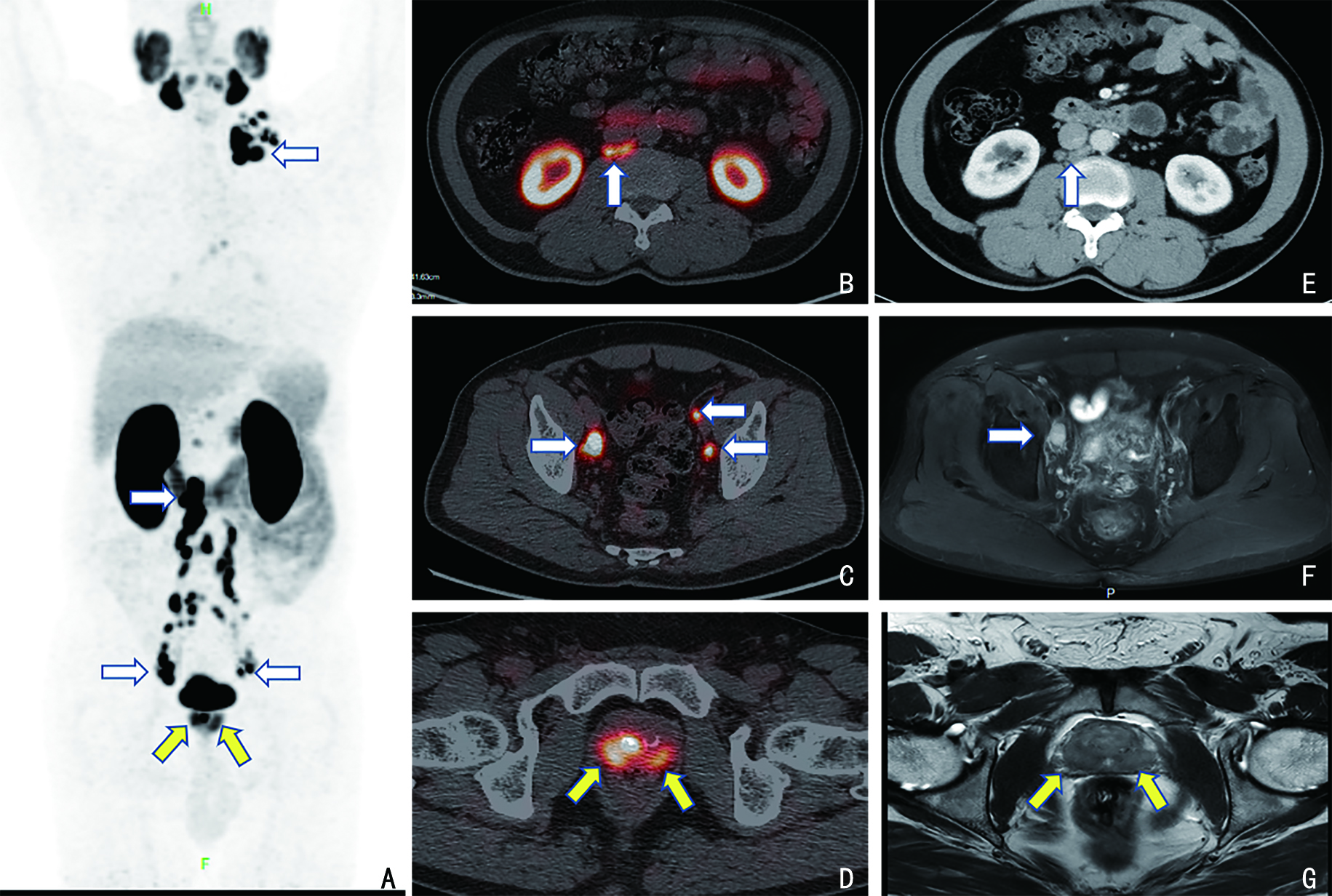
图 1 68Ga-PSMA-11 PET/CT与传统影像学检查在前列腺癌淋巴结转移中的比较
A:PSMA PET/CT MIP(maximum intensity projection)图显示前列腺癌(黄箭)及多部位淋巴结转移(白箭);B~D:PSMA PET/CT融合图像显示淋巴结(直径为3~8 mm)转移(白箭);E~G:对应层面传统影像学检查(增强CT及MRI)显示小淋巴结转移漏诊

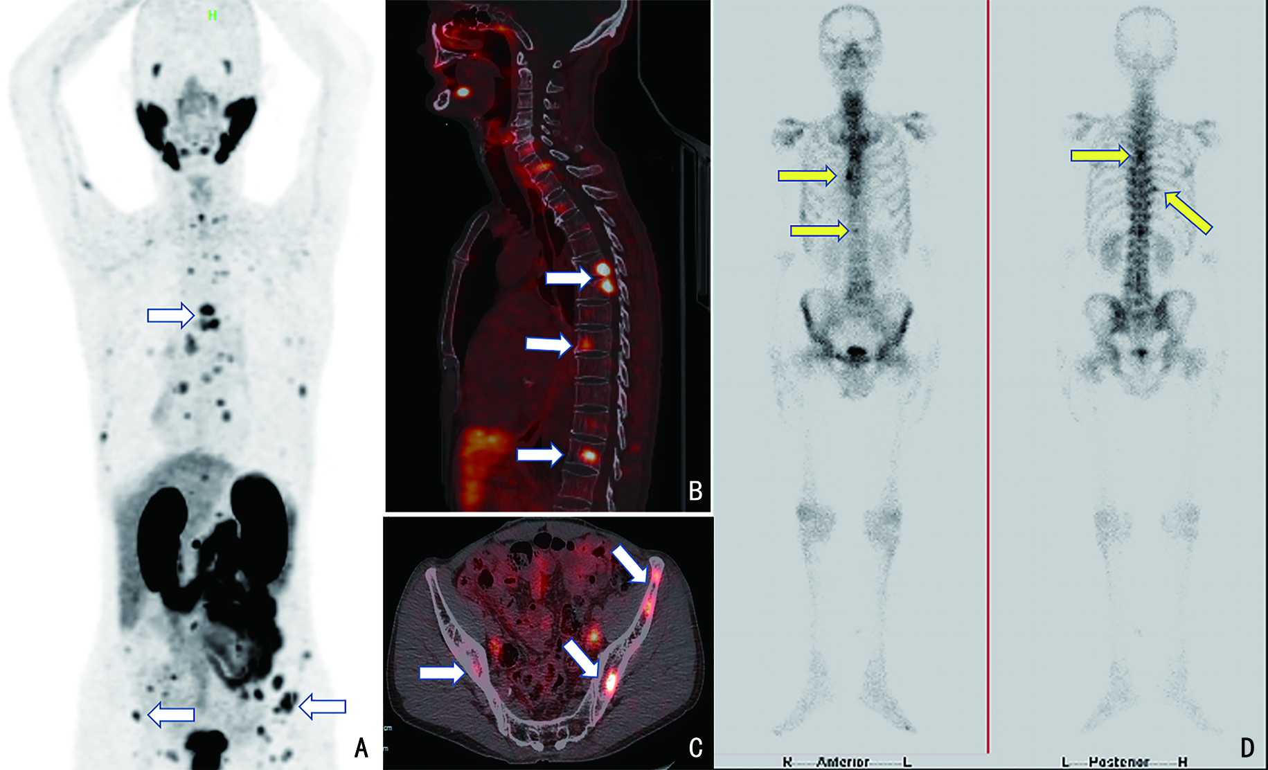
图 2 68Ga-PSMA-11 PET/CT与传统影像学检查在前列癌骨转移中的比较
A:PSMA PET/CT MIP图;B~C:PSMA PET/CT融合图像显示脊柱及骨盆多发性转移(白箭);D:传统影像学检查(骨扫描)显示骨转移(黄箭)和骨盆病灶漏诊
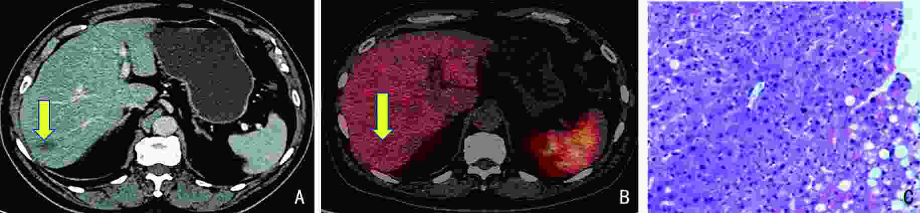
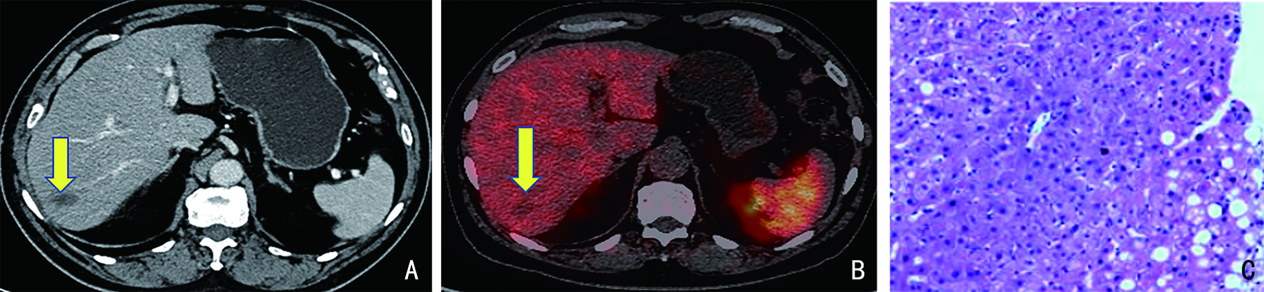
图 3 68Ga-PSMA-11 PET/CT与传统影像学检查在前列腺癌肝脏转移中的比较
A:传统影像学检查(增强CT)显示肝右叶转移可疑(黄箭);B:PSMA PET/CT融合图像显示病灶无显像剂浓聚(黄箭)并排除肝转移;C:病灶穿刺活检病理图片(H&E×100)显示肝样细胞增生伴纤维血管组织增生
表 1 63例患者行PSMA PET/CT 检查前、后的TNM分期比较
检查项目 T分期(n=63) P N分期(n=63) P M分期(n=63) P Tx T2 T3 T4 Nx N0 N1 Mx M0 M1a M1b M1c 传统影像学检查 4 23 24 12 0.414 0 44 19 0.034 0 43 2 16 2 <0.001 PSMA PET/CT 0 28 24 11 0 39 24 2 26 2 29 4  下载: 导出CSV
表 2 63例患者临床治疗方案
例(%) 治疗方案 例数(n=63) 合计 前列腺根治性切除术+盆腔淋巴结清扫术 25(39.68) 联合内分泌治疗 8(12.70) 未联合内分泌治疗 17(26.98) 前列腺根治性放疗 6(9.53) 联合内分泌治疗 5(7.94) 未联合内分泌治疗 1(1.59) 系统治疗为主 32(50.79) 内分泌治疗 25(39.69) 内分泌治疗联合化疗 2(3.17) 内分泌治疗联合盆腔放疗 2(3.17) 内分泌治疗联合前列腺局部切除 2(3.17) 内分泌治疗联合化疗及骨转移灶减症放疗 1(1.59)
下载: 导出CSV
-
[1] Kinoshita Y, Kuratsukuri K, Landas S, et al. Expression of prostate-specific membrane antigen in normal and malignant human tissues[J]. World J Surg, 2006, 30(4):628-636. doi: 10.1007/s00268-005-0544-5 [2] Lenzo NP, Meyrick D, Turner JH. Review of gallium-68 PSMA PET/CT imaging in the management of prostate cancer[J]. Diagnostics (Basel), 2018, 8(1):E16. [3] Yip KCJ, Li YL, Chen SR, et al. 68Ga-PSMA PET/CT and mpMRI for primary lymph node staging of intermediate to high-risk prostate cancer: a systematic review and meta-analysis of diagnostic test accuracy[J]. Clin Transl Imaging, 2021, 9(5):523-537. doi: 10.1007/s40336-021-00453-w [4] D'Amico AV, Whittington R, Malkowicz SB, et al. Biochemical outcome after radical prostatectomy, external beam radiation therapy, or interstitial radiation therapy for clinically localized prostate cancer[J]. JAMA, 1998, 280(11):969-974. doi: 10.1001/jama.280.11.969 [5] Buyyounouski MK, Choyke PL, McKenney JK, et al. Prostate cancer-major changes in the American Joint Committee on Cancer eighth edition cancer staging manual[J]. CA Cancer J Clin, 2017, 67(3):245-253. doi: 10.3322/caac.21391 [6] Müller J, Ferraro DA, Muehlematter UJ, et al. Clinical impact of 68Ga-PSMA-11 PET on patient management and outcome, including all patients referred for an increase in PSA level during the first year after its clinical introduction[J]. Eur J Nucl Med Mol Imaging, 2019, 46(4):889-900. doi: 10.1007/s00259-018-4203-0 [7] Donswijk ML, van Leeuwen PJ, Vegt E, et al. Clinical impact of PSMA PET/CT in primary prostate cancer compared to conventional nodal and distant staging: a retrospective single center study[J]. BMC Cancer, 2020, 20(1):723. doi: 10.1186/s12885-020-07192-7 [8] Wang XJ, Wen Q, Zhang HS, et al. Head-to-head comparison of 68Ga-PSMA-11 PET/CT and multiparametric MRI for pelvic lymph node staging prior to radical prostatectomy in patients with intermediate to high-risk prostate cancer: a meta-analysis[J]. Front Oncol, 2021, 11:737989. doi: 10.3389/fonc.2021.737989 [9] Zhao RN, Li YJ, Nie LH, et al. The meta-analysis of the effect of 68Ga-PSMA-PET/CT diagnosis of prostatic cancer compared with bone scan[J]. Medicine, 2021, 100(15):e25417. doi: 10.1097/MD.0000000000025417 [10] Morigi JJ, Anderson J, de Nunzio C, et al. Prostate specific membrane antigen positron emission tomography/computed tomography and staging high risk prostate cancer: a non-systematic review of high clinical impact literature[J]. Minerva Urol Nephrol, 2021, 73(1):32-41. [11] Wondergem M, van der Zant FM, Broos WAM, et al. 18F-DCFPyL PET/CT for primary staging in 160 high-risk prostate cancer patients; metastasis detection rate, influence on clinical management and preliminary results of treatment efficacy[J]. Eur J Nucl Med Mol Imaging, 2021, 48(2):521-531. doi: 10.1007/s00259-020-04782-2 [12] Roach PJ, Francis R, Emmett L, et al. The impact of 68Ga-PSMA PET/CT on management intent in prostate cancer: results of an Australian prospective multicenter study[J]. J Nucl Med, 2018, 59(1):82-88. doi: 10.2967/jnumed.117.197160 [13] Grubmüller B, Baltzer P, Hartenbach S, et al. PSMA ligand PET/MRI for primary prostate cancer: staging performance and clinical impact[J]. Clin Cancer Res, 2018, 24(24):6300-6307. doi: 10.1158/1078-0432.CCR-18-0768 [14] Koerber SA, Will L, Kratochwil C, et al. 68Ga-PSMA-11 PET/CT in primary and recurrent prostate carcinoma: implications for radiotherapeutic management in 121 patients[J]. J Nucl Med, 2019, 60(2):234-240. doi: 10.2967/jnumed.118.211086 [15] Fanti S, Goffin K, Hadaschik BA, et al. Consensus statements on PSMA PET/CT response assessment criteria in prostate cancer[J]. Eur J Nucl Med Mol Imaging, 2021, 48(2):469-476. doi: 10.1007/s00259-020-04934-4 [16] Kuten J, Fahoum I, Savin Z, et al. Head-to-head comparison of 68Ga-PSMA-11 with 18F-PSMA-1007 PET/CT in staging prostate cancer using histopathology and immunohistochemical analysis as a reference standard[J]. J Nucl Med, 2020, 61(4):527-532. doi: 10.2967/jnumed.119.234187 -

 点击查看大图
计量
- 文章访问数: 190
- HTML全文浏览量: 83
- PDF下载量: 41
- 被引次数: 0
